The Composition of the Systemic Arterial Blood Is Determined by the Composition of the Capillary Blood That Drains Each Alveolus
Blood that returns from the lungs to the left ventricle for distribution to the tissues comes from capillaries associated
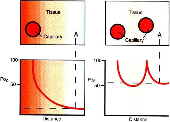
FIGURE 47-4 Effect of increasing capillary density on tissue oxygen tension (Po2).
Oxygen tension is shown as a function of the distance from the capillary. Left, Point A has Po2 of approximately 10 mm Hgz whereas, right, point A has Po2 of approximately 50 mm Hg, because it is now closer to a second tissue capillary.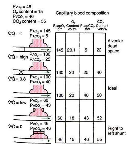
FIGURE 47-5 ■ Diagrammatic representation of an alveolus and a capillary, showing the effect of differing ventilation/perfusion (VZQ) ratios on the partial pressure and gas content of blood leaving the alveolus. In the case OfVZQ = ∞ (top), there is no bulk flow of blood past the alveolus, so capillary blood does not contribute to the arterial blood that leaves the left ventricle. See text for explanation andTable 47-1 for abbreviation definitions.
with millions of alveoli, each of which may have a slightly different VZQ ratio. The content of oxygen and carbon dioxide in blood that leaves each alveolus varies because of these differing VZQ ratios. Thus the composition of arterial blood is determined by distribution of VZQ ratios in the lung.
Lung disease accentuates VZQ mismatching because of obstruction of airways, Hooding of alveoli with exudates, and local obstructions to blood flow. This mismatching has a major effect on oxygen exchange but little effect on the exchange of carbon dioxide. In the case of oxygen, overventilation of some alveoli does not compensate for underventilation of others. Because of the shape of the oxyhemoglobin dissociation curve (see Chapter 48) the overventilated (high VZQ) alveoli with a high PAo2 cannot add enough oxygen to the blood to compensate for the deficiency that arises from the underventilated (low VZQ) alveoli with a low PAo2.
Therefore, hypoxemia occurs to varying degrees in most lung diseases. However, in contrast to oxygen, carbon dioxide is very soluble, and because its dissociation curve (see Chapter 48) is almost linear, the overventilated alveoli can compensate for those that are underventilated. For this reason, hypercarbia, also called hypercapnia (increased Paco2), rarely occurs in the presence of lung disease.As the degree of VZQ mismatching increases and oxygen exchange becomes less efficient, the difference between the average PAo2 and Pao2 increases. Normally, this alveolar-
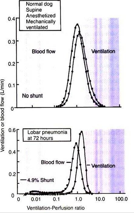
FIGURE 47-6 Distribution of Ventilation and blood flow as a function of the VentilationZperfusion (VZQ) ratio. Top, In the normal dog, most of the blood flow and ventilation are received by gas exchange units with a VZQ ratio close to 1.0. No blood flow and no ventilation are received by units with extremely high or extremely IowVZQ ratios. Bottom, In the dog with pneumonia, a considerable portion of the blood flow is received by units with IowVZQ ratios, that is, units with little ventilation.The amount of blood passing through right-to-left shunts is also increased in pneumonia. (From Wagner PD, Laravuso RB, Goldzimmer E, et al: Distributions of ventilation-perfusion ratios in dogs with normal and abnormal lungs, J Appl Physiol 38:1099, 1975.)
to-arterial oxygen tension difference (AaDo2) averages 5 to 10 mm Hg, because there is a degree of VZQ inequality even in normal lungs, and because venous blood draining the bronchial and coronary circulations mixes with the oxygenated blood draining the alveoli. The AaDo2 increases when animals are anesthetized or when they have lung disease, because many poorly ventilated regions of the lung continue to receive blood flow; that is, the number of units with low VZQ increases (see Figure 47-6).
Right-to-Left Vascular ShuntsAIIow Blood to BypassVentiIated Lung
In a right-to-left shunt, blood from the right ventricle bypasses ventilated Iungand returns to the left atrium (see Figure 47-5, bottom). Such blood does not pick up oxygen and, when it leaves the lung, has the same composition as the venous blood that entered the lung. Right-to-left shunts have a V/Q ratio of zero and are formed when alveoli are collapsed (atelectasis), are unventilated because of complete airway obstruction, or are filled with exudates, as in acute pneumonia. Right-to-left shunts can also result from complex congenital cardiac defects, such as tetralogy of Fallot, which allow blood to flow directly from the right to the left chambers of the heart, bypassing the lungs. Such large right-to-left shunts cause a major impairment of oxygen exchange. In normal animals the venous blood (low Po2) from the bronchial and coronary veins enters the oxygenated blood leaving the lungs. This is equivalent to a right-to-left shunt and constitutes 5% of cardiac output.