The Contractile Machinery in Cardiac Muscle Is Similar to That in Skeletal Muscle
Cardiac muscle, as with skeletal muscle, has a striated appearance under the light microscope (Figure 19-1). These crossstriations have the same structural basis in cardiac and skeletal muscle.
Each striated cardiac muscle fiber (muscle cell) is made up of a few hundred myofibrils. Each myofibril has a repetitive pattern of light and dark bands. The various bandsTable 19-1
IB
Sequence of Events in Contraction of Skeletal Muscle and Cardiac Muscle
Skeletal muscle
Cardiac muscle
Action potential is generated in somatic motor neuron
Acetylcholine is released
Nicotinic cholinergic receptors on muscle cell membrane are activated
Ligand-gated Na+ channels in muscle membrane open
Muscle membrane depolarizes to threshold level for formation of action potential
Action potential forms in muscle cell but does not enter other cells
Note: Skeletal muscle cells do not have slow Ca2+ channels
Action potential causes Ca2* release from sarcoplasmic reticulum; Ca2* binds to troponin
Actin,s binding sites are made available for actin-myosin cross-bridge formation
Cross-bridge cycling generates contractile force between actin and myosin filaments
Muscle contracts (brief "twitch"); Ca2* is taken up by sarcoplasmic reticulum
Muscle relaxes
Λ∕ote. Action potentials in autonomic motor neurons are not needed to initiate heartbeats
Note: Neurotransmitters are not needed to make the heart beat
/Vote: Activation of receptors is not needed. A completely isolated or denervated heart still beats
Pacemaker Na* channels spontaneously open (and K* channels close) in membranes of pacemaker cells
Pacemaker cell membranes depolarize to threshold for formation of action potential
Action potential forms in a pacemaker cell and then propagates from cell to cell throughout the whole heart
During action potential, extracellular Ca2* ("trigger" Ca2*) enters cell through "slow" Ca2* channels
Entry of extracellular trigger Ca2* causes release of more Ca2* from sarcoplasmic reticulum; Ca2* binds to troponin
Actin's binding sites are made available for actin-myosin cross-bridge formation
Cross-bridge cycling generates contractile force between actin and myosin filaments
Heart contracts (complete "beat" or "systole"); Ca2* is taken up by sarcoplasmic reticulum or pumped back out of cell into extracellular fluid
Heart relaxes
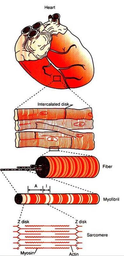
FIGURE 19-1 Under the light microscope, cardiac muscle fibers (muscle cells) are seen to be striated, similar to skeletal muscle. Electron microscopy reveals that the striations result from an orderly arrangement of actin (thin) filaments and myosin (thick) filaments into muscular subunits called sarcomeres (as shown in bottom drawing).
Sarcomeres are the structural and functional subunits of cardiac muscle, as they are in skeletal muscle. Unlike skeletal muscle fibers, however, cardiac muscle fibers often branch, and they link end to end with neighboring fibers at structures called intercalated disks. Unseen within the intercalated disks are nexi, or gap junctions, which are minute cytoplasmic channels that allow action potentials to propagate from cell to cell.and lines within a myofibril are given letter designations (A band, I band, Z disk). The alignment of these bands in adjacent myofibrils accounts for the striated appearance of the whole muscle fiber. Each repeating unit of myofibrillar bands is called a sarcomere. This name, which means “little muscle,” is apt because a single sarcomere constitutes the basic, contractile subunit of the cardiac muscle. By definition, a sarcomere extends from one Z disk to the next, a distance of approximately 0.1 mm, or 100 μm.
As in skeletal muscle, each cardiac muscle sarcomere is composed of an array of thick and thin filaments. The thin filaments are attached to the Z disks; they interdigitate with the thick filaments. The thin filaments are composed of actin molecules. The thick filaments are composed of myosin molecules. In the presence of adenosine triphosphate (ATP) and calcium ions (Ca2^), myosin and actin interact in a series of steps called the cross-bridge cycle, which results in contraction and force generation in each sarcomere and therefore in the whole muscle cell (for details, see Figures 1-3, 1-4» 1-5, and 6-5).