» The Elbow, Forearm, and Carpus
The elbow joint projects onto the ventral ends of the fourth and fifth ribs. The olecranon, the medial and lateral epicondyles of the humerus, and the robust collateral ligaments are all easily palpable and provide the necessary orientation for joint puncture.
This is performed from the lateral aspect with the needle directed between the lateral epicondyle and the olecranon to enter a considerable pouch of the capsule within the deep olecranon fossa.Long Bone Fractures: Food animals experience a high incidence of fractures, mainly of the long bones. Among the long bone fractures, those of the metacarpus (III/IV) and metatarsus account for nearly half.
The ulna is complete but slender, and it is the massive radius that bears the weight. As always the subcutaneous medial border of the radius marks the division between the cranial extensor and caudal flexor muscle groups (Fig. 30.3). The ulna is palpable only at its extremities, the olecranon and lateral styloid process. In most subjects the forearm inclines mediodistally to the carpus while the foot angles laterally, producing a "knock-kneed" stance. Although straight limbs are preferred, this inward bulging of the carpus does not appear to be a disadvantage.
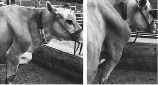
FIG. 30.1 “Wing shoulder” in a 6-year-old Jersey cow.
The proximal row of the carpal skeleton comprises radial, intermediate, and ulnar carpal bones. The upper and lower borders of the accessory bone provide rough guides to the levels of the antebrachiocarpal and midcarpal joints. The distal row consists of only two bones: fused second and third carpals and the fourth carpal (see Fig. 2.48). In theory, movement is possible at all three levels, but most occurs between the forearm and carpus, a moderate amount takes place at the middle joint, and next to none occurs at the carpometacarpal level.
Movements other than flexion and extension are largely prevented by the many ligaments, of which the collateral pair is most important. The cavities of the two distal joints always communicate; occasionally all three do so. Puncture is possible at the proximal and middle levels and is obviously most easily performed when the joint is flexed.Irregularities of the palmar aspect of the carpal bones are covered and smoothed by the thick fibrous layer of the joint capsule (palmar carpal ligament), which combines with the accessory bone and flexor retinaculum to enclose the carpal canal. The joint capsule also bends dorsally with deep fascia to form the extensor retinaculum that binds the extensor tendons in place. An inconstant bursa between this retinaculum and the skin occasionally enlarges to form an unsightly but painless blemish (hygroma).
Hygromas of the carpus and tarsus result from repeated trauma or inadequate bedding and are manifested as fluid accumulation in subcutaneous tissue. These may require procedures such as drainage or surgical resection.
Only the digital extensors and flexors among the muscles of the forearm merit notice. The common digital extensor has two bellies: the larger medial one extends its tendon of insertion to the medial digit, and the smaller lateral belly has a tendon that splits at the fetlock to insert on both digits. The two tendons share a synovial sheath where they descend over the carpus. The lateral digital extensor comports itself like the medial belly of the common extensor (Fig. 30.2). The superficial digital flexor also possesses two bellies. The three bellies of the deep flexor give rise to a stout tendon that receives synovial protection during its passage through the carpal canal, while the superficial one remains outside the flexor retinaculum. Both are protected by long synovial sheaths that extend beyond the carpus into the cannon where the tendons merge. All these tendons receive further discussion later.
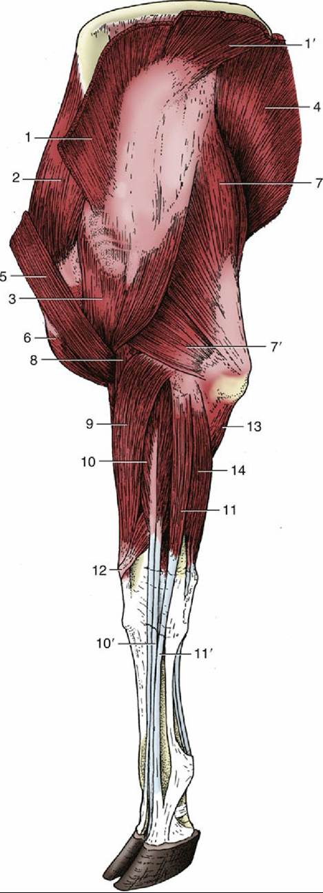
FIG.
30.2 Muscles of the bovine forelimb, lateral view. 1 and 1', Trapezius; 2, supraspinatus; 3, deltoideus; 4, latissimus dorsi; 5, brachiocephalicus; 6, biceps; 7 and 7', long and lateral heads of triceps, respectively; 8, brachialis; 9, extensor carpi radialis; 10, common digital extensor; 10', tendon of lateral belly; 11 and 11', lateral digital extensor and its tendon, respectively; 12, extensor carpi obliquus; 13, ulnar head of deep digital flexor; 14, ulnaris lateralis.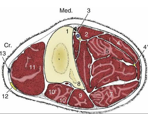
FIG. 30.3 Transverse section of the middle of the bovine left forearm. Cr., Cranial; Med., medial; 1, radius; 2, flexor carpi radialis; 3, median vessels and nerve; 4, flexor carpi ulnaris; 4', ulnar nerve; 5, superficial digital flexor; 6, deep digital flexor; 7, ulnaris lateralis; 8, ulna; 9, lateral digital extensor; 10 and 10', common digital extensor; 11, extensor carpi radialis; 12, superficial branch of radial nerve; 13, cephalic vein.