THE ENDOCRINE GLANDS
The paired thyroid glands (Figure 37-37Z5) of the chicken are reddish-brown, oval, and about 10 mm long
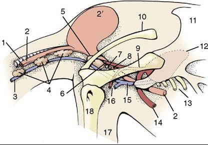
Figure 37-37 Junction of neck and trunk as viewed from the right, semischematic.
Cranial is to the left. 1, Trachea; 2, esophagus; 2', crop; 3, right jugular v.; 4, thymus; 5, thyroid gland; 6, right common carotid a.; 7, parathyroid glands; 8, ultimobranchial gland; 9, right brachiocephalic a.; 10, clavicle; 11, sternum; 12, position of heart; 13, sternal ribs; 14, descending aorta; 15, right cranial vena cava; 16, subclavian a. and v.; 17, wing; 18, humerus.and 5 mm wide. In the budgerigar, in which thyroid disease is a major problem in iodine-deficient areas, they are paler and only 2 to 3 mm long and 1 to 2 mm wide. The thyroid glands are located in the thoracic inlet, caudal to the crop and closely related to the common carotid artery, the trachea, the jugular vein, and vagus nerve (which accompanies the vein)—indeed they lie just cranial to where these vessels are joined by the subclavian vessels (Figure 37-37/16). Their color distinguishes them from the neighboring rather similar but pale thymic lobes.
The parathyroid glands (Figure 37-37/7), two or three on each side, are minute (1- to 3-mm) yellowish-brown structures immediately caudal to the thyroid gland to which one may be attached. They become enlarged (increased parathyroid hormone production) when the diet is deficient in calcium, which leads to decalcification of the bones. In African grey parrots (Psittacus erithacus) there is a specific problem in which calcium fails to be mobilized from the skeleton despite a dietary deficiency. In this situation the bird will die of hypocalcemia and much enlarged parathyroids will be found at necropsy.
The even more minute pink ultimobranchial glands (Figure 37-37/8) lie next to the parathyroids.
The adrenal glands (Figure 37-31/5') are yellowish brown, oval or triangular, and about 13 mm long and 8 mm wide. Each lies at the cranial pole of the corresponding kidney, related ventrally to the ovary (or epididymis). There is no distinct separation of cortex and medulla.
The hypophysis (or pituitary gland) (Figure 37-38/7) is attached below the diencephalon and occupies the hypophysial fossa in the base of the skull. It resembles that of mammals in its division and formation.