The esophagus (or gullet) conveys food from the pharynx to the stomach.
This relatively narrow tube begins dorsal to the cricoid cartilage of the larynx and follows the trachea down the neck, at first inclining to the left but regaining a median position above the trachea before or shortly after entering the thorax (Fig.
3.29). Within the thorax it runs in the mediastinum (p. 147); continuing beyond the tracheal bifurcation, it passes over the heart before penetrating the esophageal hiatus of the diaphragm. It then makes its way over the dorsal border of the liver to join the stomach at the cardia. It thus has cervical, thoracic, and abdominal portions, although the last is very short.The cervical part of the esophagus runs within the visceral space of the neck, related to the subvertebral muscles dorsally and the left side of the trachea medioventrally (see Fig. 3.29) and accompanied by the left common carotid artery and vagosympathetic and recurrent laryngeal nerves.
The thoracic part crosses to the right of the aortic arch. Caudally, its dorsal and ventral borders are followed by the respective vagal trunks into which fibers of the right and left vagus nerves are regrouped.
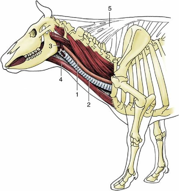
FIG. 3.29 Lateral view of the bovine neck. In the midneck the esophagus lies on the left dorsolateral aspect of the trachea. 1, Esophagus; 2, trachea; 3, pharyngeal musculature; 4, sternocephalicus muscle; 5, nuchal ligament.
The structural pattern of the esophagus is similar to that of the rest of the alimentary canal. The outer coat is a loose connective tissue (adventitia) in the neck, but this is largely replaced by serosa* in the thorax and abdomen. The muscle is striated at the origin of the esophagus, but in some species (e.g., cat, pig, and horse) the striated muscle is replaced by smooth muscle at some point within the thorax.
Both layers of the muscles are spiral, and they wind in opposite directions in the first part of the esophagus. Closer to the stomach the outer coat becomes more longitudinal and the inner one more circular (Fig. 3.30). There is considerable interlacing of muscle bundles between the two layers. Although not proven morphologically, a number of esophageal sphincters are suggested by functional studies. They include a cranial sphincter, probably provided by fibers of the cricopharyngeus muscle and possibly others within the thorax, where the passage of food tends to be delayed. Although a thickening suggestive of a sphincter occurs at the junction of the esophagus with the stomach, the flow of food is actually more impeded immediately in front of the diaphragm.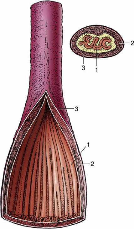
FIG. 3.30 Semischematic drawing of the structure of the esophagus, sectioned longitudinally and transversely. 1, Mucosa; 2, muscular layer (longitudinal and circular); 3, adventitia.
The inner part of the wall is divided between submucosa and mucosa by a fenestrated muscularis mucosae, usually more prominent in the thoracic esophagus (Fig. 3.31B). It helps throw the lining of the empty organ into longitudinal folds. The surface epithelium is generally stratified squamous, and the degree of keratinization and thickness reflects the diet of the species, as illustrated in Fig. 3.31. In comparison with the goat esophagus, the canine esophagus has many submucosal mucussecreting tubuloacinar glands. The boundary between esophageal and gastric epithelia is sharp and may be displaced to either side of the cardia. In humans, prolonged or repeated exposure to gastric juice (e.g., heartburn) may provoke transformation of the stratified epithelium of the lower esophagus into the columnar gastric variety.
The esophagus receives its innervation from the sympathetic and vagus nerves, including the recurrent laryngeal branches.
The vagal supply is the more important. The striated muscle arises from the mesoderm of the pharyngeal arches and is under control of the general visceral motor neurons of the vagus, whereas the smooth muscle portions are under direct control of the intrinsic nervous system and indirect control of the autonomic nervous system. A myenteric plexus extends the length of the esophagus.The blood supply from various local arteries presents no features of special interest.
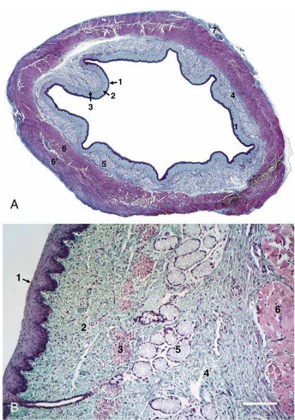
FIG. 3.31 Esophagus, in the dog (A) stained with Masson's trichrome showing collagen as green and
cellular components as purple red and goat (B) stained with hematoxylin and eosin (70?) showing four
main tunics: 1, mucosa; 2, submucosa; 3, muscular layer and 4, adventitia. Mucosal layer is composed of 1' stratified squamous epithelium; 1”, lamina propria (connective tissue); and 1”’, lamina muscularis
mucosae (smooth muscles). Submucosa contains 5, mucus-secreting tubuloacinar glands in the connective tissue and the muscle layer is arranged in 3', inner layer; and 3”, outer layer.