THE EYE
The eyeball resembles that of the globular mammalian one. The general structure is globular, although the shape may differ, especially in its anterior part, which may be flat, globose, or tubular, depending on the species (Figure 37-39).
The eyeball almost fills the orbit, leaving little room for movement; however, the long neck and mobile occipitoatlantal joint compensate for this.The lower lid is the larger and more movable. The third eyelid has a stiffened edge; being translucent, it does not seem to impair vision when drawn across the cornea. The secretions of the lacrimal gland and the
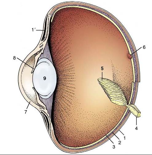
Figure 37-39 Section through the eyeball, schematic. 1, Sclera; 1', ring of scleral ossicles; 2, choroid; 3, retina; 4, optic nerve; 5, pecten; 6, fovea centralis; 7, cornea; 8, iris; 9, lens.
deep gland of the third eyelid leave the conjunctival sac through two puncta that lead to a spacious nasolacrimal duct. The upper punctum is surprisingly large.
The cornea is thin and strongly curved. Its small diameter belies the enormous eyeball to which it belongs. The sclera is reinforced by a layer of cartilage transformed into a ring of ossicles near the cornea (Figure 37-39Z1'). No tapetum lucidum is present. The iris of the chicken is yellow-brown but turns slightly paler during the laying period. It surrounds a round pupil that can rapidly change in size through the action of the striated sphincter and dilator muscles. Even so, the avian iris is surprisingly unresponsive to light. In most other species the iris is darker, ranging from brown to black, although it can be bright yellow in owls. In African grey parrots the grey iris of the juvenile becomes yellow at maturity. In cockatoos the female has a red to brown iris, and the male has dark brown to black. The retina is devoid of blood vessels. It displays a remarkable outgrowth (pecten; Figure 37-39Z5) over the optic disc. This is a black, pleated ridge that projects into the vitreous; rich in blood vessels, it is thought to play a role in the nutrition of the retina. The extraocular muscles are similar to those of mammals, although a retractor bulbi is lacking.