» The Eye
The orbital rim projects above the surrounding surfaces. The orbital cavity is capacious, although reduced Ventrorostrally by the fragile, thin-walled swelling of the lacrimal bulla into which the maxillary sinus extends.
The orbital axes diverge upward, outward, and forward and together subtend an angle of approximately 120 degrees. It is therefore clear that, as is usual in ungulates, the field of monocular vision is large and that of binocular vision is small.The eyelids are supported by dense fibrous plates or "tarsi." The skin adheres tightly over the orbicularis muscle but is loose elsewhere, leading to a furrowed lid when the eye is open. The lashes are long and are more densely spread on the upper lid. The muscles of the lids include the frontalis, which extends from the forehead into the upper lid, and the malaris, which radiates from the lower lid onto the face. These are supplied by the facial nerve, mainly through the auriculopalpebral nerve. The levator, supplied as always by the oculomotor nerve, remains active in facial paralysis, which mitigates the effects of this injury.
The conjunctiva contains considerable scattered lymphoid accumulations in its palpebral part. The usual glands are present within the eyelids. The largest, the tarsal (meibomian) glands, occupy the deeper layers of the tarsi; they may be visible through the conjunctiva of the everted lid.
Infectious bovine keratoconjunctivitis (pink eye) is caused by Moraxella bovis. This disease causes significant economic losses to the dairy industry through treatment and management costs. The initial mucous and pus discharge is followed by a corneal ulcer.
The medial corner of the palpebral opening forms a bay containing the fleshy lacrimal caruncle. The third eyelid covers a variable part of the bulb. The supporting cartilage sinks medial to the eyeball, where it is associated with superficial and deep accessory lacrimal glands.
Only a small part of the third eyelid is normally visible. A larger part is brought into view when the eyeball is withdrawn or pressed into its socket; this displaces the retrobulbar fat, which in turn pushes the cartilage and therefore the fold outward.The lobulated, bipartite lacrimal gland lies dorsolaterally on the eyeball. It drains by numerous ducts of varying caliber into the upper conjunctival fornix. The tears collect by the lacrimal caruncle before entering the slitlike puncta lacrimalia that lead to the lacrimal sac. The sac lies within a depression of the cranial part of the orbital wall. It tapers to the nasolacrimal duct, which first traverses the maxillary sinus and then runs on the lateral nasal wall to discharge within the nasal vestibule.
The extrinsic muscles, which exhibit no especially notable features, are shown schematically in Fig. 9.19.
The eyeball is small in relation to the orbit. The sclera is thin and locally obtains a bluish tinge from the dark underlying choroid. Some pigmentation is common, especially toward the junction with the cornea, and tends to increase with age. The cornea is ovoid, and its pointed end is lateral. It is rather thick, especially toward its margin.
The bovine pupil is widened from side to side when constricted but becomes circular on dilation. Its upper and lower margins are broken by irregular projections, the iridic granules, which are smaller than in the horse; they are more prominent along the upper margin. The ciliary muscles are poorly developed, and the capacity for accommodation is limited accordingly. The vascular and choroidocapillary layers of the choroid are separated in the caudal part of the bulb by the brilliantly colored reflective tapetum (Fig. 25.21). The tapetum is triangular, and its base is directly above the optic disk. Its peripheral parts are most colorful and display an array of metallic blues and greens, while the area close to the optic disk is reddish, especially in the calf.
Ophthalmoscopic examination of the tapetum reveals scattered dark flecks, where capillaries enter, and larger vessels, which appear as red lines. Four pairs of arteries and veins radiate in cruciate fashion from the optic disk, which is lateroventral to the posterior pole of the eye. The dorsal vein is especially large and is entwined by a spiraling artery. A clear spot in the center of the disk indicates the vestige of the hyaloid artery; as would be expected, the remnant is more obvious in the newborn calf. The macula of the retina consists of two rather ill-defined parts: a rounded area placed dorsolateral to the optic disk is concerned with binocular vision, and a horizontal strip below the tapetum is concerned with monocular vision. Their extents are suggested by their relatively poor vascularization.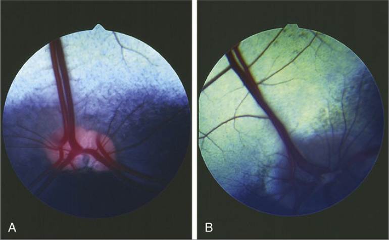
FIG. 25.21 (A) Fundus of eye of cow. (B) Fundus of eye of goat.
Evisceration of the orbit is sometimes performed under local anesthesia. The anesthetic technique, though simple, is exacting because it requires the deposit of anesthetic solution deep in the orbit, precisely by the single foramen (orbitorotundum) through which emerge the nerves that supply the structures within the periorbita. The nerves are thus blocked where bundled together before dispersing to their scattered destinations. Movement of the eyelids may be prohibited in the usual way—that is, by blockage of the palpebral branch of the facial nerve where it crosses the zygomatic arch (Fig. 25.6/3).