The Facial Nerve (VII)
The facial nerve is sometimes known as the intermediofacial nerve, a term that indicates its composite nature. The intermediate component is a visceral one with sensory (including gustatory) and motor (parasympathetic) functions; the facial component is the nerve of the second pharyngeal arch whose main distribution is to the muscles of the face (mimetic musculature).
The facial nerve arises close to the vestibulocochlear nerve at the lateral extremity of the trapezoid body (Fig. 8.20/VII and VIII), and the two nerves run within common meningeal covering to the internal acoustic meatus of the petrous temporal bone. The facial nerve enters the facial canal within the bone that leads, via a sharp caudal convexity ("genu"), to the stylomastoid foramen, where the nerve appears at the surface of the skull. The facial nerve contains the appropriately named geniculate ganglion at the corner of the convexity within the facial canal. With the exception of a small branch to the stapedius muscle, the branches of the facial nerve detached within the facial canal represent the intermediate (visceral) component and those detached after leaving the bone the motor component (Fig. 8.70/1).
Within the facial canal, the greater petrosal nerve branches from the main nerve at the level of the ganglion and emerges through an independent foramen. It initially contains only parasympathetic axons but is shortly joined by sympathetic fibers to form a composite autonomic nerve, the nerve of the pterygoid canal. This nerve runs through the pterygoid canal to reach the pterygopalatine ganglion within the pterygopalatine fossa (Fig. 8.71/7 and 11). The nerve of the pterygoid canal is discussed more fully later (p. 313). The stapedial nerve, which arises next within the canal, is motor to the stapedius muscle of the middle ear. The next branch, the chorda tympani (Fig.
8.71/13), crosses the tympanic cavity to emerge at the petrotympanic fissure, after which it converges on and becomes incorporated within the lingual branch of the mandibular nerve (p. 338).After the facial nerve emerges from the stylomastoid foramen, its first branches are the internal and caudal auricular nerves, which supply muscles of the external ear and several branches to hyoid muscles, including the caudal belly of the digastricus. The main trunk enters the face by turning around the mandible, where it is first contained between the masseter and the parotid gland. It divides at about this level (although there are species differences) into three terminal branches, the auriculopalpebral nerve and the dorsal and ventral buccal nerves.
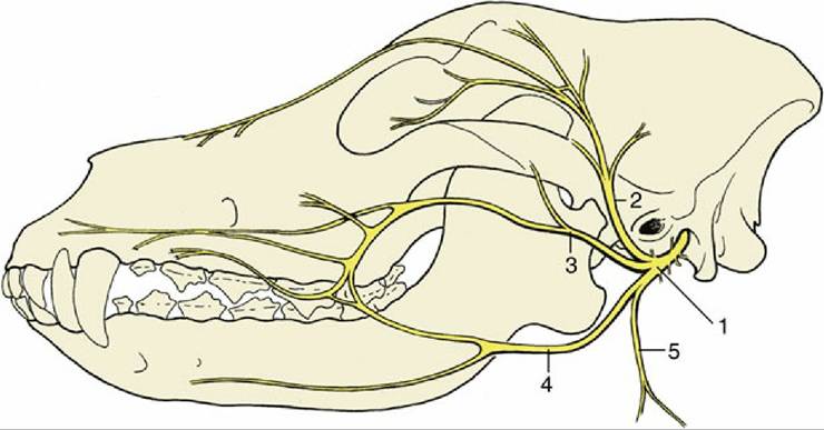
FIG. 8.70 Distribution pattern of the facial nerve (n.) of the dog. 1, Facial n.; 2, auriculopalpebral n.; 3,
dorsal buccal branch; 4, ventral buccal branch; 5, cervical branch.
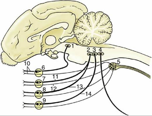
FIG. 8.71 Schematic representation of the autonomic innervation of structures of the head. 1, Parasympathetic oculomotor nucleus (III); 2, parasympathetic facial nucleus (VII); 3, parasympathetic glossopharyngeal nucleus; 4, parasympathetic vagus nucleus; 5, cranial cervical ganglion; 6, ciliary ganglion; 7, pterygopalatine ganglion; 8, mandibular ganglion; 9, otic ganglion; 10, short ciliary nerves; 11, greater petrosal nerve; 12, deep petrosal nerve; 13, chorda tympani; 14, tympanic plexus, short petrosal nerve.
In some species the auriculopalpebral nerve (Fig. 8.70/2) is detached before the main trunk reaches the face, and it is then less vulnerable to injury from superficial trauma to the side of the head. It crosses the zygomatic arch, traveling toward the space between the upper eyelid and external ear, before dividing into branches that supply the muscles of the eyelids (excluding the levator palpebrae superioris) and the auricular muscles in front of the external ear.
The dorsal buccal branch (Fig. 8.70/3), which may take the form of a group of divergent branches, crosses the masseter en route to the muzzle.
In some species the ventral buccal branch (Fig. 8.70/4) may take a path similar to that of the dorsal branch at a slightly more ventral level, but in others it takes a divergent course, first running within the intermandibular space before entering the face with the parotid duct and facial vessels, where they cross the mandible in front of the masseter. Together, the buccal branches supply the muscles of the cheek, lips, and nostrils. Their peripheral branches join with those of the trigeminal nerve at various levels, and many of the smaller trunks combine motor (facial) and sensory (trigeminal) fibers.
The effects of injury or disease clearly depend on the site of the lesion. Lesions that are situated more centrally, which tend to have more serious origins, affect the whole facial field and lead to loss of secretory activity by the lacrimal and salivary (except the parotid) glands in addition to muscular paralysis. Lesions involving the main trunk near its exit from the bone paralyze the entire facial musculature, whereas more peripheral lesions may spare some function, depending on their site and the specific and individual variations in the branching pattern. Lesions confined to the auriculopalpebral nerve produce drooping of the external ear and narrowing of the palpebral fissure with inability to close the eye. Damage to the buccal branches may paralyze the muscles of the lips and cheeks, allowing food to collect in the oral vestibule. It may also lead to deformation of the muzzle, which is made asymmetrical by the unopposed activity of the muscles on the sound side. The alteration in appearance is not always very striking, and the uninjured side, toward which the muzzle is pulled, may sometimes appear to have the more distorted aspect. The distortion tends to be more pronounced in the horse and sheep than in other domestic species. It is important to be aware that in unilateral facial spasm, seen occasionally in the dog, the nose may be drawn toward the affected side.
The auriculopalpebral nerve is sometimes blocked to facilitate examination of the eye.