THE FLEXOR MUSCLES
The flexor muscles comprise the biceps brachii and braşchialis. Although largely under cover of the brachioceşphalicus, the belly of the biceps is palpable as it lies against the cranial face of the humerus.
The biceps takes origin from the supraglenoid tubercle of the scapula by means of a short, broad, and largely fibroşcartilaginous tendon that is molded on the intertuberşcular groove. The (intertubercular) bursa that protects the tendon spreads from the groove onto the cranial aspect of the humerus; it may be a cause of shoulder lameness when inflamed. The bursa may be reached, certainly if overdistended, by inserting a needle between the muscle and the bone, slightly above the level of the deltoid tuberosity, and then directing it proximally (Figure 23-11/3).The biceps inserts mainly on the radial tuberosity, but a branch of the attachment passes beneath the medial collateral ligament to the adjoining parts of the radius and ulna. A more important peculiarity is the existence within its belly of a fibrous strand (internal tendon; Figure 23-10/5') that joins the tendons of origin and insertion; a part splits away to emerge on the surface and blend more distally with the epimysium of the extensor carpi radialis. The bridging band, known as the lacertus fibrosus, is easily found as a firm structure crossing the flexor aspect of the elbow
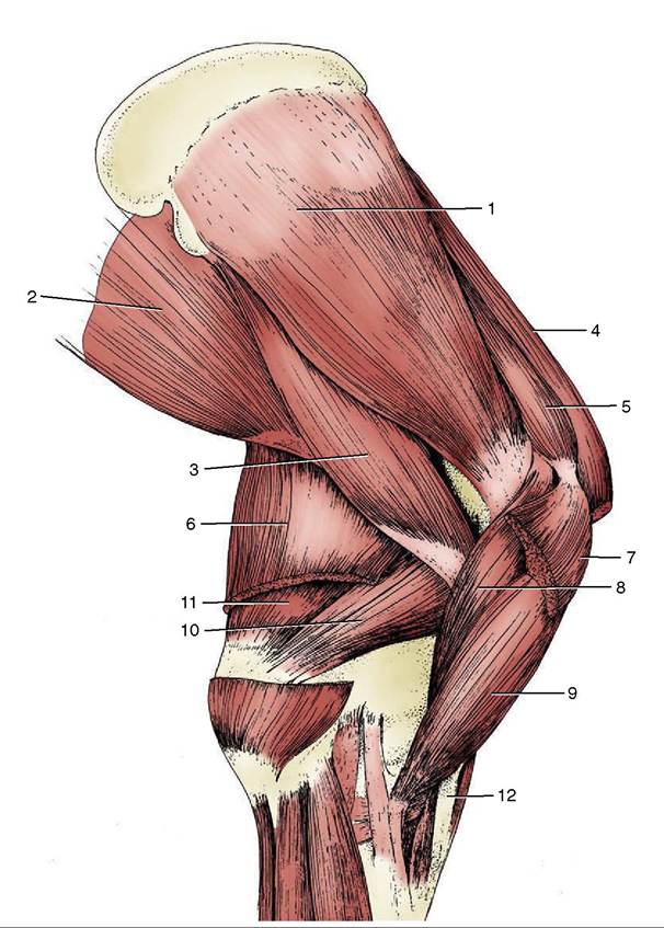
Figure 23-8 Muscles on the medial surface of the right shoulder and arm. 1, Subscapularis; 2, latissimus dorsi; 3, teres major; 4, subclavius; 5, supraspinatus; 6, tensor fasciae antebrachii; 7, deep pectoral; 8, coracobrachialis; 9, biceps; 10, medial head of triceps; 11, long head of triceps; 12, lacertus fibrosus.
(Figure 23-10/5"; Figure 23-8/72). It is taut in the standing animal but slackens as the joint is flexed.
The internal tendon and the lacertus help maintain the carpal joint in extension when the biceps resists collapse of the shoulder under the weight of the trunk (Figure 23-38, AJ2,6).The biceps is a fixator and, potentially, an extensor of the shoulder; the construction and form of the tendon of origin suggest its particular fitness for the first task. Although it is regarded as the most important flexor of the elbow, the fibrous arrangements imply that its passive role may also be more significant at this joint. Recent work has furnished a more satisfactory explanaştion of the unusual structure and enigmatic role of the equine biceps brachii than has been available hitherto. Calculation has shown that the force necessary for the rapid protraction of the forelimb that is effected at the faster gaits is beyond the power of the available muscle as conventionally assessed. It is now suggested that the central tendon allied to the bipennate construction of the muscle enables it to store energy when stretched during the support phase of the stride and that this energy is later very rapidly released to accelerate the forward movement of the limb. Its nerve supply comes from the musculocutaneous nerve.
The brachialis is purely fleshy and crosses only one joint, the elbow. It arises from the caudoproximal part of the humerus, winds laterally within a spiral groove, and then crosses the flexor aspect of the elbow to insert
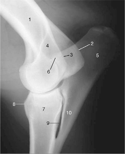
Figure 23-9 Lateral radiograph of an elbow joint. 1, Humerus; 2, medial epicondyle; 3, lateral epicondyle; 4, olecşranon fossa; 5, olecranon; 6, anconeal process of olecranon; 7, radius; 8, radial tuberosity; 9, interosseous space; 10, ulna.
on the Craniomedial part of the proximal radius (Figure 23-12/5). Proximally, the muscle is covered by the triceps, but its distal part is superficial and may be palşpated. The brachialis is purely an elbow flexor. It is supplied by the musculocutaneous nerve with, rather surprisingly, a contribution from the radial nerve.