The General Plan of the Animal Body
All farm animals are vertebrates, and as such they have a vertebral column. The body (with the exception of some of the internal organs) exhibits bilateral symmetry. This means that the right and left sides of the body are mirror images of each other.
similar right and left structures are called paired structures, such as a pair of gloves that are similar but not interchangeable. Most unpaired structures are on or near the median plane, and of course, only one of each unpaired structure exists in any given animal. The tongue, trachea, vertebral column, and heart are examples of unpaired structures. The ribs, limbs, eyes, and most muscles are paired structures.Wherever organs are expected to be in more- or-less constant motion and must glide past one another without friction (e.g., the beating heart and moving gut), a serosal cavity is present. The simple squamous epithelium lining various body cavities is also called mesothelium, and the cavities have within them only a scant amount of fluid to facilitate free movement of the tissues. The diaphragm divides the embryonic body cavity into a thoracic cavity and the abdominopelvic cavity. Each of these are further subdivided.
The thoracic cavity contains the pericardial sac, which surrounds the heart, and two pleural sacs, which surround the two lungs. These sacs are formed by a serous membrane, the pleura, a layer of simple squamous epithelium with underlying connective tissue, moistened with the small amount of fluid within the cavity of the sac.
The abdominopelvic cavity is somewhat arbitrarily divided into the abdominal and pelvic cavities. The abdominal cavity contains the kidneys, most of the digestive organs, and a variable amount of the internal reproductive organs in both sexes. The pelvic cavity contains the terminal part of the digestive system (the rectum) and all of the internal portions of the urogenital system not found in the abdominal cavity.
The abdominal and pelvic cavities are continuous with one another, and the brim of the pelvis marks the transition. The serous membrane that surrounds the abdominal viscera and part of the pelvic viscera is called peritoneum.A transverse section through the abdominal cavity illustrates the general plan of the body as a tube (the digestive tract and its derivatives) within a tube (the body wall) (Fig. 1-8). Normally there are few air-filled spaces in the animal body except in the respiratory system and the ear. However, for the sake of clarity, many illustrations show a considerable separation between structures that in the animal body are actually in contact.
The layers of the body wall and the layers of the digestive tract show a striking similarity, although in reverse order. Layers of the body
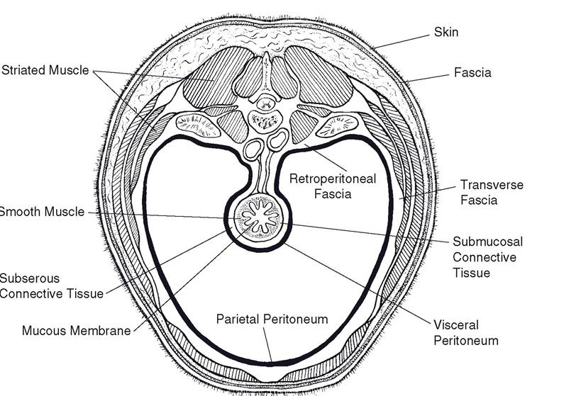
Figure 1-8. Cross-section of the body wall and digestive tract.
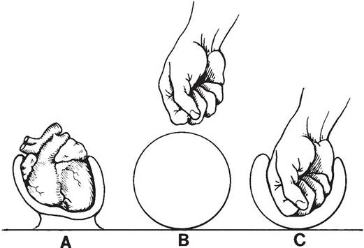
Figure 1-9. A) Invagination of serous membrane to form outer (parietal) and inner (visceral) layers. B and C) This is similar to a fist pushed into a balloon.
wall from outside inward are (1) epithelium (epidermis of the skin), (2) connective tissue (dermis and fascia), (3) muscle (striated), (4) connective tissue (transverse fascia), and (5) mesothelium (parietal peritoneum). The layers of the gut wall from outside inward are (1) mesothelium (visceral peritoneum), (2) connective tissue (subserous connective tissue), (3) muscle (smooth), (4) connective tissue (submucosa), and (5) epithelium (mucous membrane) (Fig. 1-8).
The serous membranes mentioned previously (pericardium, pleura, and peritoneum) are all derivatives of the lining of the celomic cavity of the embryo. Each serous membrane forms a continuous sac that is usually empty except for a small amount of serous (watery) fluid. In other words, no viscera are found inside any of the serous sacs, although most viscera are covered by at least one layer of a serous membrane.
A simple analogy is that of pushing one’s fist into a partially inflated balloon. The fist is never actually within the balloon proper, but still it is surrounded by a portion of the balloon (Fig. 1-9).The part of the serous membrane covering a viscus is called the visceral serous membrane (visceral pericardium, visceral pleura, and visceral peritoneum). The serous membrane lining a body cavity is called the parietal serous membrane (parietal pericardium, parietal pleura, and parietal peritoneum). The continuity of each serous sac is maintained by connecting layers of serous membrane that extend from the visceral layer of each serous membrane to the parietal layer of the same serous membrane. The names of these connecting layers of serous membranes are based on the specific areas they connect, and they are discussed in some detail along with the relevant systems later in this book.