Microscopic Anatomy: Animal Cells and Tissues
All living things, both plants and animals, are constructed of small units called cells. The simplest animals, such as the ameba, consist of a single cell that is capable of performing all functions commonly associated with life.
These functions include growth (increase in size), metabolism (use of food), response to stimuli (such as moving toward light), contraction (shortening in one direction), and reproduction (development of new individuals of the same species).A typical cell consists of three main parts, the cytoplasm, the nucleus, and the cell membrane (Fig. 1-2). Detailed structure of the individual cell is described in chapter 2. Tissues are discussed in this chapter.
In complex animals, certain cells specialize in one or more the functions of the animal
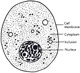
Figure 1-2. A cell as seen with a light microscope.
body. A group of specialized cells is a tissue. For example, cells that specialize in conducting impulses make up nerve tissue. cells that specialize in holding structures together make up connective tissue. Various tissues are associated in functional groups called organs. The stomach is an organ that functions in digestion of food. A group of organs that participate in a common enterprise make up a system. The stomach, liver, pancreas, and intestines are all part of the digestive system.
The primary types of tissues include (1) epithelial tissues, which cover the surface of the body, line body cavities, and form glands; (2) connective tissues, which support and bind other tissues together and from which, in the case of bone marrow, the formed elements of the blood are derived; (3) muscle tissues, which specialize in contracting; and (4) nervous tissues, which conduct impulses from one part of the body to another.
Epithelial Tissues
In general the epithelial tissues are classified as simple (composed of a single layer) or stratified (many-layered). Each of these types is further subdivided according to the shape of the individual cells within it (Fig. 1-3). simple epithelium includes squamous (platelike) cells, cuboidal (cubic) cells, columnar (cylindrical) cells, and pseudostratified columnar cells.
Simple squamous epithelium consists of thin, platelike cells. They are much expanded in two directions but have little thickness. The edges are joined somewhat like mosaic tile covering a floor. A layer of simple squamous epithelium has little tensile strength and is found only as a covering layer for stronger tissues. simple squamous epithelium is found where a smooth surface is required to reduce friction. The coverings of viscera and the linings of body cavities and blood vessels are all composed of simple squamous epithelium.
Cuboidal epithelial cells are approximately equal in all dimensions. They are found in some ducts and in passageways in the kidneys.
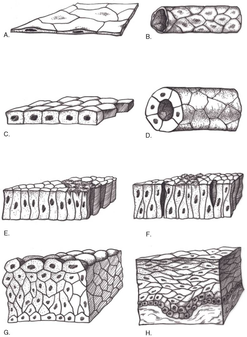
Figure 1-3. Primary types of epithelial tissues. A) Simple squamous. B) Simple squamous in tubular arrangement. C) Simple cuboidal. D) Simple cuboidal arranged as a duct. E) Simple columnar. F) Pseudostratified columnar with cilia. G) Transitional. H) Stratified squamous.
The active tissue of many glands is composed of cuboidal cells.
Columnar epithelial cells are cylindrical. They are arranged somewhat like the cells in a honeycomb. some columnar cells have whiplike projections called cilia extending from the free extremity.
Pseudostratified columnar epithelium is composed of columnar cells. However, they vary in length, giving the appearance of more than one layer or stratum. This type of epithelium is found in the upper respiratory tract, where the lining cells are ciliated.
Stratified epithelium consists of more than one layer of epithelial cells and includes stratified squamous, stratified columnar, and transitional epithelia.
Stratified squamous epithelium forms the outer layer of the skin and the lining of the first part of the digestive tract as far as the stomach. In ruminants, stratified squamous epithelium also lines the forestomach (rumen, reticulum, and omasum). stratified squamous epithelium is the thickest and toughest of the epithelia, consisting of many layers of cells. From deep to superficial, these layers include the basal layer (stratum basale), the parabasal layer (stratum spinosum), intermediate layer (stratum gran ulosum), and superfi cial layer (stratum corneum). The deepest layer, the stratum basale, contains the actively growing and multiplying cells. These cells are somewhat cuboidal, but as they are pushed toward the surface, away from the blood supply of the underlying tissues, they become flattened, tough, and lifeless and are constantly in the process of peeling off. This layer of cornified (keratinized) dead cells becomes very thick in areas subjected to friction. calluses are formed in this manner.
Stratifi ed columnar epithelium is composed of more than one layer of columnar cells and is found lining part of the pharynx and salivary ducts.
Transitional epithelium lines the portions of the urinary system that are subjected to stretching. These areas include the urinary bladder and ureters. Transitional epithelium can pile up many cells thick when the bladder is small and empty and stretch out to a single layer when completely filled.
Glandular epithelial cells are specialized for secretion or excretion. Secretion is the release from the gland cell of a substance that has been synthesized by the cell and that usually affects other cells in other parts of the body. Excretion is the expulsion of waste products.
Glands may be classified either as endocrine glands (glands without ducts, which empty their secretory products directly into the bloodstream), or as exocrine glands (glands that empty their secretory products on an epithelial surface, usually by means of ducts).
The endocrine glands are an important part of the control mechanisms of the body, because they produce special chemicals known as hormones. The endocrine glands are discussed in Chapter 12. Hormones carried to all parts of the body by the blood constitute the humoral control of the body. Humoral control and nervous control are the two mechanisms maintaining homeokinesis, also called homeostasis, a relatively stable but constantly changing state of the body. Humoral responses to stimuli from the environment (both external and internal) are slower and longer acting than responses generated by way of the nervous system. The nervous system is described in some detail in Chapters 9 and 10.
Collectively, the endocrine glands constitute the endocrine system, which is studied in endocrinology. However, exocrine glands are scattered throughout many systems and are discussed along with the systems to which they belong, such as the digestive, urogenital, and respiratory systems.
According to their morphologic classification (Fig. 1-4), a gland is simple if the duct does not branch and compound if it does. If the secretory portion forms a tubelike structure, it is called tubular; if the secretory portion resembles a grape or hollow ball, it is called alveolar or acinar (the terms are used interchangeably). A combination of tubular and alveolar secretory structures produces a tubuloalveolar gland.
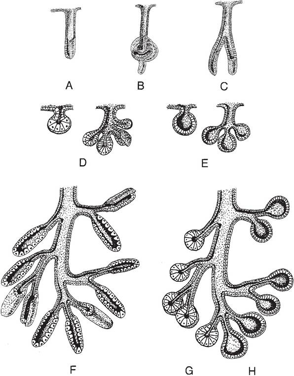
Figure 1-4. Types of exocrine glands and comparison of simple and compound glands. A) Simple tubular gland. B) Simple coiled tubular gland. C) Simple branched tubular gland. D & E) Simple acinar/alveolar glands and simple branched acinar/alveolar glands. F) Compound tubular gland. G & H) Compound acinar/alveolar glands. Compound tubuloacinar/tubuloalveolar glands consist of either a mixture of tubular and acinar/alveolar secretory units or tubular secretory units “capped” by acini or alveoli.
(Reprinted with permission of Wiley-Blackwell from Eurell, J.A. and Frappier, B.L. DellmannS Textbook of Veterinary Histology. 6th ed. Ames, IA: Blackwell Publishing Professional, 2006.)Compound glands often are subdivided grossly into lobes, which in turn may be further subdivided into lobules. Hence, the connective tissue partitions (called septa) are classified as interlobar septa if they separate lobes and as interlobular septa if they separate lobules. Similar terminology may be applied to ducts draining lobes or lobules of glands, that is, interlobar ducts and interlobular ducts, respectively.
Another classification of glands is based on the manner in which their cells elaborate their secretion. By this classification, the most common type is the merocrine gland. Merocrine glands pass their secretory products through the cell wall without any appreciable loss of cytoplasm or noticeable damage to the cell membrane. The holocrine gland is the least common type. After the cell fills with secretory material, the entire holocrine gland cell discharges to the lumen of the gland to constitute the secretion. sebaceous glands associated with hair follicles of the skin are the most common holocrine glands. An intermediate form of secretion in which a small amount of cytoplasm and cell membrane is lost with the secretion is sometimes described for the prostate and some sweat glands. Such glands are called apocrine glands.
Connective Tissues
Connective tissues, as the name implies, serve to connect other tissues. They give form and strength to many organs and often provide protection and leverage. connective tissues include elastic tissue, collagenous (white fibrous) tissue, reticular (netlike) tissue, adipose (fat) tissue, cartilage, and bone.
Elastic tissue contains kinked fibers that tend to regain their original shape after being stretched. This tissue is found in the ligamentum nuchae, a strong band that helps to support the head, particularly in horses and cattle.
Elastic tissue also is found in the abdominal tunic, in the ligamenta flava of the spinal canal, in elastic arteries, and mixed with other tissues wherever elasticity is needed.Collagenous (white fibrous) tissue is found throughout the body in various forms. Individual cells (fibroblasts) produce long proteinaceous fibers of collagen, which have remarkable tensile strength. These fibers may be arranged in regular repeating units, or laid down in a more random, irregular arrangement.
In dense regular connective tissue (Fig. 15), the fibers are arranged in parallel bundles, forming cords or bands of considerable strength. These are the tendons, which connect muscles to bones, and the ligaments, which connect bones to bones.
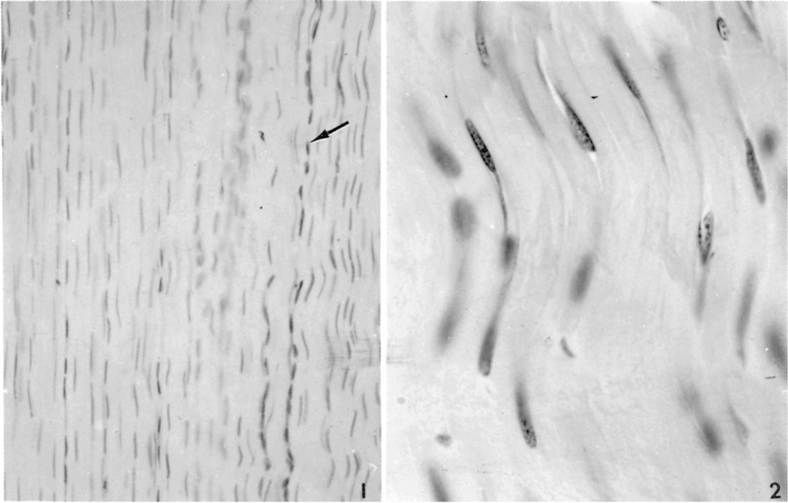
Figure 1-5. Longitudinal section through a tendon showing the histological appearance of dense regular connective tissue. (Left) notice the line of nuclei (arrow), indicating the loose connective tissue surrounding blood vessels and nerves. Hematoxylin and eosin stain, ?226. At higher power (right), spindle-shaped fibroblasts can be seen among collagen fibers. Hematoxylin and eosin stain, ?660. (Reprinted with permission of Wiley-Blackwell from Dellmann, H.D. and Brown, E.M. Textbook of Veterinary Histology. 2nd ed. Philadelphia: Lea & Febiger, 1981.)
The fibers of dense irregular connective tissue are arranged in a thick mat, with fibers running in all directions. The dermis of the skin, which may be tanned to make leather, consists of dense irregular connective tissue. This forms a strong covering that resists tearing and yet is flexible enough to move with the surface of the body.
Areolar (loose) connective tissue (Plate I) is found throughout the body wherever protective cushioning and flexibility are needed. For example, blood vessels are surrounded by a sheath of areolar connective tissue, which permits the vessels to move and yet protects them.
Beneath the dermis is a layer of loosely arranged areolar connective tissue fibers that attaches the skin to underlying muscles. This attachment is flexible enough to permit movement of the skin. it also permits the formation of a thick layer of fat between the skin and underlying muscles. Whenever the skin is adherent to bony prominences because of a lack of areolar tissue, the skin will not move, and no layer of fat can form. This feature is seen in beef cattle that have ties; in this case, the skin over the back shows large dimples where fat cannot fill in because the skin is adherent to the vertebrae.
Reticular connective tissue consists of fine fibrils and cells. Reticular tissue makes up part of the framework of endocrine and lymphatic organs.
Adipose tissue (fat) forms when connective tissue cells called adipocytes store fat as inclusions within the cytoplasm of the cell. As more fat is stored, the cell eventually becomes so filled with fat that the nucleus is pushed to one side of the cell, which, as a result, becomes spherical (Plate I). Most fat in the animal body is white, although it may have a yellow tinge in horses and some breeds of dairy cattle because of carotenoids in the feed.
In contrast to this white fat, a small amount of brown fat may be found in domestic mammals, hibernating mammals, rodents, and human infants. The brown fat is found between the scapulae, in the axillae, in the mediastinum, and in association with mesenteries in the abdomen. Brown fat apparently generates heat to protect young mammals and hibernating mammals from extreme cold.
Cartilage is a special type of connective tissue that is firmer than fibrous tissue but not as hard as bone. The nature of cartilage is due to the structure of the intercellular material found between the chondrocytes (cartilage cells). The three types of cartilage described are hyaline, elastic, and fibrous.
Hyaline cartilage is the glasslike covering of bones within joints. This type of cartilage forms a smooth surface that reduces friction, so that one bone easily glides over another. The actively growing areas near the ends of long bones also consist of hyaline cartilage. Elastic cartilage consists of a mixture of cartilage substance and elastic fibers. This type of cartilage gives shape and rigidity to the external ear. Fibrocartilage consists of a mixture of cartilage and collagenous fibers, which forms a semielastic cushion of great strength. The intervertebral disks between the bodies of adjacent vertebrae are composed of fibrocartilage.
Bone is produced by bone-forming cells called osteoblasts. These cells produce osteoid tissue, which later becomes calcified to form bone. The bone may be arranged in the form of spicules (small spikes) and flat plates, forming a spongelike network called cancellous bone or spongy bone. Alternatively, it may be laid down in the form of laminated cylinders (Haversian or osteonal systems), closely packed together to form compact bone (Plate I).
Blood. Blood consists of a fluid matrix (liquid portion), the plasma, a variety of cells (Plate II), proteins, monosaccharides (simple sugars), products of fat degradation, and other circulating nutrients, wastes, electrolytes, and chemical intermediates of cellular metabolism. It is sometimes considered to be a connective tissue because of the origin of some of its components.
Red blood cells (RBCs) are also called erythrocytes. In most domestic mammals they are nonnucleated biconcave disks that contain the protein hemoglobin. The main function of the RBCs is to carry hemoglobin. Hemoglobin in turn has the primary function of carrying oxygen from the lungs to all tissues of the animal. At the tissue level, oxygen is released to the cells, while carbon dioxide, which is produced by the cells, diffuses into the blood to be carried back to the lungs, where it can be eliminated during breathing. Anemia is a reduction in the concentration of functional RBCs in the blood. It can result from a loss of red cells (as in hemorrhage), insufficient RBC production, or inappropriate or premature degradation of the red cells.
White cells (also called leukocytes) are one of the body’s first lines of defense against infection. They include agranulocytes and granulocytes. Agranulocytes are of two kinds: monocytes, large cells that engulf and destroy foreign particles, and lymphocytes, which usually are smaller and are associated with immune responses. An excess of agranulocytes tends to be associated with chronic types of diseases.
Granulocytes (polymorphonuclear leukocytes) are of three types and are described according to their affinity for different stains. Granules in neutrophils stain indifferently; basophils have dark-staining granules when stained with common blood stains; and eosinophils have red-staining granules. Blood platelets (thrombocytes) are small, irregularly shaped cellular fragments that are associated with the clotting of the blood. Mammalian platelets lack a nucleus.
Plasma is the fluid part of unclotted blood. Plasma is particularly useful as a substitute for blood in transfusions because the proteins in it give it the same osmotic pressure as blood. Plasma therefore will not escape from blood vessels as readily as a salt solution.
Serum is the supernatant fluid that remains after a clot forms and incorporates the cellular components of blood. It is similar to plasma but lacks most of the clotting factors. serum is sometimes administered for prevention and treatment of diseases because it contains the antibody fractions of the blood.
Muscle Tissue
The three types of muscle tissue are skeletal, smooth, and cardiac (Plate I; Fig. 1-6). Both skeletal and cardiac muscle cells consist of
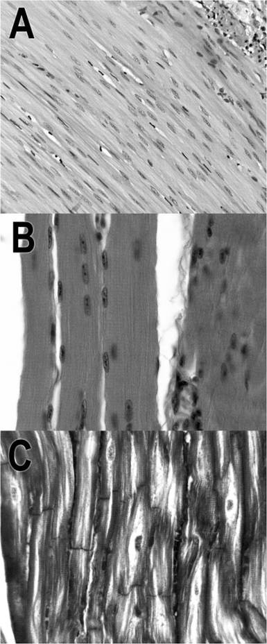
Figure 1-6. Types of muscle tissue. A) Smooth muscle. B) Skeletal muscle. C) Cardiac muscle. (Courtesy of Sandra Pitcaithley, DVM.)
fibers that under the microscope show characteristic cross-striations, so both are classified as striated muscle. Smooth muscle cells lack distinct cross-striations.
Each skeletal muscle cell must have its own nerve supply, and when stimulated, the whole fiber contracts. This is the all-or-none law of muscle contraction. However, the force of contraction depends on the state of the fiber at any one moment. For example, is it already fatigued? is it warmed up? is it stretched? striated skeletal muscle tissue plus some connective tissue makes up the flesh of meat-producing animals.
Smooth muscle cells are spindle-shaped cells that contain one centrally located nucleus per cell. smooth muscle is found in the walls of the digestive tract, in the walls of blood vessels, and in the walls of urinary and reproductive organs. These cells contract more slowly than skeletal muscle and in response to a variety of stimuli, although they are not under voluntary control.
Cardiac muscle is also known as involuntary striated muscle because it is not usually under conscious control, yet it does have crossstriations. The heart muscle is composed of a complex branched arrangement of cardiac muscle cells. Modified muscle cells called Purkinje fibers conduct impulses within the heart, much as nerve fibers do in other parts of the body.
Nervous Tissue
The essential cell of nervous tissue is the neuron (nerve cell). The neuron consists of a nerve cell body and two or more nerve processes (nerve fibers). The processes are called axons if they conduct impulses away from the cell body and dendrites if they conduct impulses toward the cell body (Fig. 1-7).
Bundles of axons in the spinal cord are called tracts, and those in the periphery are called nerves. A nerve fiber may be covered by a myelin sheath, a specialized wrapping created by supportive cells called Schwann cells in nerves or by oligodendrocytes within the brain and spinal cord.
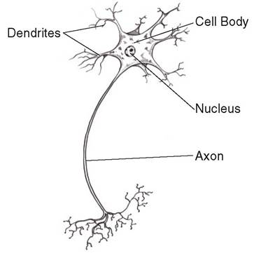
Figure 1-7. A typical motor neuron.
The special connective tissues of nervous tissue are called neuroglia and are found only in the central nervous system. outside the central nervous system, in addition to the schwann cells, ordinary white fibrous tissue serves as the major protective covering for the nerves.