THE GIRDLE MUSCLES
The same muscles join the limb to the trunk as in other species, but there are certain differences in detail. The trapezius arises from the dorsal midline, extending almost from the poll to beyond the withers.
Both cervi-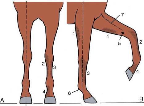
Figure 23-1 Desirable conformation and autonomous zones of cutaneous innervation of the forelimb. A, Cranial view; a vertical broken line dropped from the point of the shoulder bisects the limb. B, Right lateral view; a vertical line dropped from the tuberosity of the scapular spine bisects the limb down to the fetlock. The autonomous zones represent skin areas innervated solely by the nerves below. 1, Caudal cutaneous antebrachial nerve (ulnar); 2, medial cutaneous antebrachial nerve (musculocutaneous); 3, ulnar nerve; 4, median nerve; 5, chestnut; 6, ergot; 7, cephalic vein.
cal and thoracic parts insert on the spine of the scapula, and when they act in unison, they raise this bone against the trunk. The cervical part acting alone swings the scapula forward, which advances the limb, whereas the thoracic part acting alone swings it in the opposite direction. Both parts may be visibly outlined through the skin when contracted. The nerve supply is the accessory nerve.
The brachiocephalicus (Figure 23—3/4) arises from the mastoid region of the skull and inserts on a ridge of the humerus that extends distally from the deltoid tuberosity. It is intimately joined in the neck to the omotrans- versarius (Figure 23—3/6), which takes origin from the transverse processes of the more cranial cervical vertebrae and ends at the clavicular intersection that divides the brachiocephalicus into cervical (cleidomastoideus) and brachial (cleidobrachialis) parts. The dorsal edge of the omotransversarius is connected to the trapezius by the superficial fascia.
The ventral edge of the brachiocephalicus is clearly delineated, at least in its cranial half, as it forms the upper margin of the jugular groove (see Figure 18-38, B).The muscle is broadest over the shoulder joint, where it covers the origin of the biceps and the insertions of the supraspinatus and infraspinatus. Bilateral action flexes the neck ventrally when that part is free to move. Unilateral action in the same circumstances bends the neck toward the active side; when the neck is fixed and
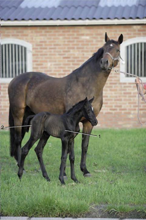
Figure 23-2 This photograph of a 10-day-old foal with its dam illustrates the proportions of the limbs and trunk that account for the "leggy" appearance of the young foal. 1, Flaccid long and medial heads of triceps; 2, "poverty" line between biceps femoris and semitendinosus.
it is the limb that is free, unilateral action advances the limb. The innervation is shared by the accessory, cervical, and axillary nerves.
The latissimus dorsi (Figure 23-3/13) arises from the supraspinous ligament and thoracolumbar fascia and converges to an insertion on the teres tuberosity of the humerus. The cranial strip covers the caudal angle of the scapula and holds it against the trunk. This muscle is commonly described as a retractor of the limb and thus is an antagonist of the brachiocephalicus; in fact, its most important role, especially in draft animals, may be to pull the trunk forward onto an advanced limb. It is supplied by the thoracodorsal nerve.
The superficial layer of girdle muscles is completed by the two superficial pectoral muscles. The cranial pectoralis descendens arises from the manubrium and divides its insertion between the humerus and fascia of
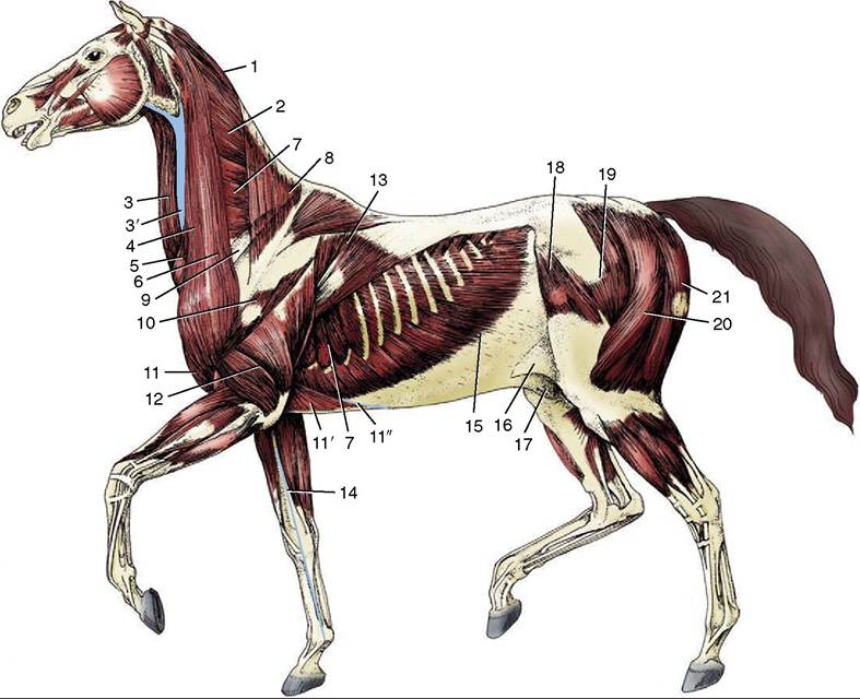
Figure 23-3 The superficial muscles and veins. The cutaneous muscles except for the cutaneous colli have been removed.
1, Rhomboideus; 2, splenius; 3, sternocephalicus; 3', jugular vein; 4, brachiocephalicus; 5, cutaneous colli; 6, omotransversarius; 7, serratus ventralis; 8, trapezius; 9, subclavius; 10, deltoideus; 11, pectoralis descendens; 11', pectoralis ascendens; 11", superficial thoracic vein; 12, triceps; 13, latissimus dorsi; 14, cephalic vein; 15, external abdominal oblique; 16, stump of cutaneous trunci forming flank fold; 17, sheath; 18, tensor fasciae latae; 19, gluteus superficialis; 20, biceps femoris; 21, semitendinosus.the arm (Figure 23-4/4). It is well developed and clearly outlined in life; a median groove separates it from its contralateral fellow. The lateral groove that marks its boundary with the brachiocephalicus is occupied by the cephalic vein. It is primarily an adductor.
The caudal pectoralis transversus (Figure 23-4/5) arises from the cranial sternebrae and inserts into the fascia over the medial aspect of the upper part of the forearm. The transverse course of its fibers makes it clear that it is essentially an adductor, which is a term that embraces the lateral shifting of the trunk toward a previously abducted limb. Both superficial pectoral muscles are supplied by pectoral branches of the brachial plexus.
Although the rhomboideus lies deep to the trapezius, it may, when contracted, form a visible surface feature. Its origin from the nuchal and supraspinous ligaments extends between the second cervical and seventh thoracic vertebrae. The entire muscle inserts on the deep face and dorsal edge of the scapular cartilage (Figure 23-5/4). Although it serves to raise the scapula, the course of the thoracic fascicles enables them to rotate the bone so that the ventral angle is carried caudally. The innervation is by dorsal branches of caudal cervical nerves.
The serratus ventralis (Figure 23-5/1) is very strong, both actively because of its extent and bulk and passively because it is covered and intershot by stout connective tissue sheets.
The origin spreads from the fourth cervical vertebra to the tenth rib. The insertion is confined to the scapular cartilage and to two triangular areas on the adjacent part of the medial surface of the scapula. The dominant function of the serratus is support of the trunk. However, the cervical and thoracic parts each have an additional (and antagonistic) function in rotating the scapula. The cervical part rotates the bone so that the ventral angle is carried caudally, thus retracting the limb; contraction of the thoracic part advances this angle and thus the limb. The serratus ventralis is supplied by the long thoracic nerve.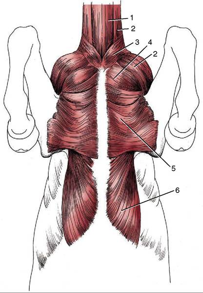
Figure 23-4 Muscles on the ventral surface of the thorax. 1, Sternocephalicus; 2, brachiocephalicus; 3, cutaneous colli; 4, pectoralis descendens; 5, pectoralis transversus; 6, pectoralis profundus.
The pectoralis profundus has a widespread origin from the caudal part of the sternum and adjacent area of the abdominal floor (Figure 23-5/5). The fascicles converge, and the muscle thickens as it passes craniolat- erally to a restricted insertion on the greater and lesser tubercles of the humerus. The relative heights of the origin and insertion suggest that the deep pectoral muscle may assist the serratus in supporting the weight of the trunk; the catastrophic results of rupture of the serratus suggest the limited effectiveness in this capacity (Figure 23-5, B). Its foremost uses are probably adduction, retraction of the limb when this is free to move, and advancement of the trunk onto an advanced and fixed limb. It is supplied by pectoral nerves.
The subclavius (Figure 23-5/2), to the front of the deep pectoral, takes origin from the cranial part of the sternum. It then bends dorsally to follow the cranial surface of the supraspinatus, over which it tapers to an extended insertion on the epimysium. Its presence along the leading edge of the scapula helps smooth the transition from the narrow neck to the greater breadth between the shoulders. The actions of the subclavius complement those of the deep pectoral (of which it was formerly regarded as a part). It too is supplied by pectoral nerves.