The Gluteal Muscles
The superficial and deep fasciae of the croup and thigh continue the corresponding coverings of the loins. The deep fascia detaches various septa that find anchorage on the pelvic girdle and the caudal edge of the sacrosciatic ligament after passing between certain muscles.
The most substantial of these separate the gluteus superficialis and biceps femoris, the biceps and semitendinosus, and the semitendinosus and semimembranosus, thus molding the muscles so that their individual contours are often clearly visible through the skin, especially in animals in "hard" training and when the muscles are contracted. The inner surface of this fascia itself, including the sides of the septa, gives origin to many fascicles of the muscles it covers.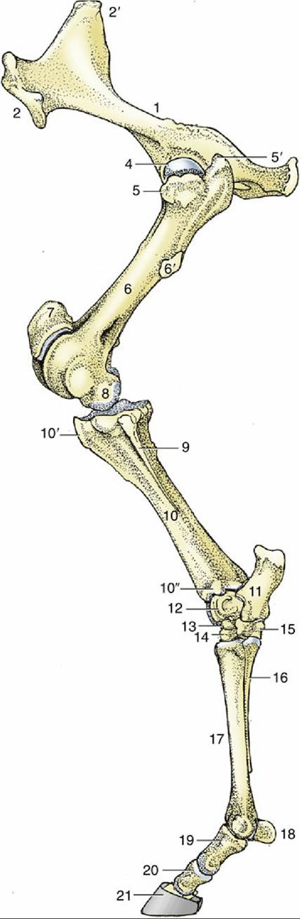
FIG. 24.1 The skeleton of the left hindlimb, lateral view. 1, Hip bone (os coxae); 2, coxal tuber; 2', sacral tuber; 3, ischial tuber; 4, head of femur; 5 and 5', cranial and caudal parts of greater trochanter, respectively; 6, femur; 6', third trochanter; 7, patella; 8, femoral condyle; 9, fibula; 10, tibia; 10', tibial tuberosity; 10", lateral malleolus; 11, calcaneus; 12, talus; 13, central tarsal; 14, third tarsal; 15, fourth
tarsal; 16, metatarsal IV (lateral splint bone); 17, metatarsal III (cannon bone); 18, proximal sesamoid bones; 19, 20, and 21, proximal, middle, and distal phalanges, respectively—the last within the hoof.
The tensor fasciae latae (Fig. 24.2/3) radiates from its origin on the coxal tuber to end by a broad aponeurosis (fascia lata) that inserts on the patella, the lateral patellar ligament, and the cranial border of the tibia. The cranial border of the fleshy part is related to the subiliac lymph nodes. The tensor is a flexor of the hip that helps to advance the limb during the swing phase of the stride.
It is supplied by the cranial gluteal nerve.The gluteus superficialis lies between the tensor and biceps (Fig. 24.2/4), with separate origins from the coxal tuber and the gluteal fascia but a single insertion on the third trochanter. Occasionally, the third trochanter is broken off and pulled dorsally by the attaching muscle. The gluteus superficialis is potentially a flexor of the hip and abductor of the thigh. Its two parts are separately supplied by the cranial and caudal gluteal nerves.
The gluteus medius is a muscle of exceptional size and power (Fig. 24.2B/2'). Its wide origin spreads from a depression scooped in the surface of the longissimus dorsi, over the coxal tuber and iliac wing, to the sacrum and adjacent part of the sacrosciatic ligament. The principal insertion is to the caudal part of the greater trochanter, but a deep division—the gluteus accessorius—has a separate aponeurotic attachment to the intertrochanteric line of the femur. This aponeurosis passes over the cranial part of the trochanter, where its passage is eased by the interposition of a synovial (trochanteric) bursa. This bursa may become inflamed. Horses so afflicted obtain relief by standing with the affected limb somewhat abducted and, when moving, by adopting an oblique doglike gait, swinging the limb in an arc.
This muscle is primarily an extensor of the hip, but it has a secondary use as an abductor of the thigh. Its association with the longissimus dorsi makes it an effective participant in rearing. It is supplied by the cranial gluteal nerve.
The gluteus profundus lies deep to the caudal part of the gluteus medius. It arises from and around the ischial spine and passes more or less transversely to insert on the cranial part of the greater trochanter. An abductor of the thigh, it is supplied by the cranial gluteal nerve (Table 24.1).
» TABLE 24.1
Gluteal Muscles
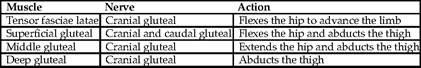
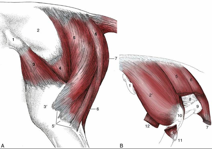
FIG. 24.2 (A) Muscles of the croup and thigh, lateral view. (B) Croup muscles, resected to expose the ischial tuber; lateral view. 1, Coxal tuber; 2, deep gluteal fascia; 2', gluteus medius; 3, tensor fasciae latae;
3', fascia lata; 4, gluteus superficialis; 5, vertebral head of biceps; 5', the three distal divisions of the biceps; 6, semitendinosus; 7, semimembranosus; 8, sacrosciatic ligament; 9, ischial tuber; 10, caudal part of greater trochanter; 11, third trochanter; 12, stump of rectus femoris.