» The Heart
The heart lies in the ventral part of the middle mediastinum, directly cranial to the diaphragm and largely covered by the forelimbs (Fig. 20.1). It forms an irregular and laterally compressed cone.
The larger part of the heart lies left of the median plane and is so disposed that the axis slopes caudoventrally and to the left (Fig. 20.3). The heart of a Thoroughbred obviously is conspicuously larger, both relatively and absolutely, than that of other horses of comparable body weight. The difference is mostly inherited and partly conditioned by training and affects the topography. Most commonly the heart extends between the planes of the second to sixth intercostal spaces, which places the apex directly caudal to the level of the point of the elbow. The cranial margin is strongly curved and is arranged with its upper part vertical, and its lower part follows the dorsal surface of the sternum. The caudal border, though sinuous in profile, is more or less upright (Fig. 20.3). The flattened lateral surfaces are related through the pericardium to the mediastinal surfaces of the lungs, except where the cardiac notches allow direct contact, greater on the left side, with the thoracic wall. A strong sternopericardiac ligament attaches the pericardium to the sternum, and this, with the anchorage of the great vessels, limits the displacement allowed to the heart. A slight shift, however, does occur with the movement of the diaphragm.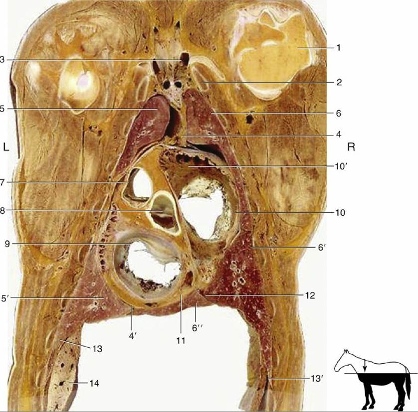
FIG. 20.8 Dorsal section of the thorax at the level of the atrioventricular valves. 1, Head of humerus; 2, first rib; 3, formation of cranial vena cava; 4 and 4', cranial and caudal mediastinum, respectively; 5 and 5', cranial and caudal lobes of the left lung, respectively; 6, 6', and 6", cranial, caudal, and accessory lobes of
the right lung, respectively; 7, pulmonary valve; 8, aortic valve; 9, left atrioventricular valve; 10, right atrioventricular valve; 10', right auricle; 11, coronary sinus; 12, plica venae cavae; 13, diaphragm; 13', costodiaphragmatic recess; 14, part of the liver; L, left side; R, right side.
Apart from the general form, there is little of significance to distinguish the heart of the horse.
Mention should be made, however, of two features of the aortic and pulmonary valves, especially of the former. The cusps commonly develop nodules at the free margins, and these can be quite striking in older animals. In addition, fenestrations may appear in the middle region of the cusps. Neither development appears to have much, if any, functional significance. The puncta maxima, the sites at which the valve sounds are most clearly heard, do not correspond exactly to the projections of the openings on the chest wall.Auscultation: The left atrioventricular valve is auscultated to most advantage in the fifth intercostal space, a little caudodorsal to the point of the elbow; the aortic valve at a somewhat higher level in the fourth space; and the pulmonary valve lower within the third space — all of course on the left side. The right atrioventricular valve is best heard in the lower parts of the third and fourth right intercostal spaces.
It is important to remember that the skeletal topography is not always easy to appreciate in practice. It may be more useful to remember that the puncta lie within a band of a few centimeters' depth about midway between the horizontal planes that intersect the points of the shoulder and the elbow. Within this band the punctum maximum of the left atrioventricular valve is at the intersection of the vertical line that falls a couple of fingerbreadths behind the point of the elbow. The approach to the other valves follows from the relative positions indicated and requires the introduction of the stethoscope between the limb and the chest wall.
The coronary arteries share the supply of the heart wall in more equal fashion than in many other species because the right one ends by descending within the right (subsinuosal) interventricular groove (see Fig. 7.19/2').