The Heart
The heart is placed asymmetrically, 60% or more being to the left of the midline, and extends from the second intercostal space (or following rib) to the fifth space. It thus lies mainly under cover of the limbs in an animal standing square.
The base lies in the plane of the last costochondral joint and the apex opposite the sixth cartilage, a few centimeters above the sternum; its long axis inclines somewhat caudally and to the left. Direct contact with the thoracic walls is restricted to the areas described with the lungs. The upright caudal border is related to the diaphragm and, through this, to the reticulum and liver; the sloping cranial border is related to the thymus in the young. The relations of the base include the trachea and principal bronchi, the pulmonary vessels, and lymph nodes (Fig. 27.5).The bovine heart is constructed according to the general mammalian plan. The right atrium receives a left azygous vein, by way of the coronary sinus. It occasionally retains communication with the left atrium through an open foramen ovale; this is usually only probe patent and without significance. Two ossicles are found in the connective tissue related to the cusps of the aortic valve; they are not unique to cattle, as often supposed, but do appear to develop precociously in this species. The left coronary artery is dominant, with the right one restricted to a circumflex course. It is worth mentioning that the isthmus of the aorta (the segment between the origin of the brachiocephalic trunk and the junction with the ductus arteriosus) is greatly constricted in the newborn calf, which is an appearance that may falsely suggest the aorta arising from the right ventricle. The heart attains usual proportions in neonates within a few days of the birth.
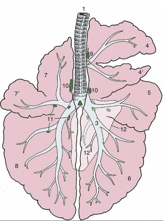
FIG.
27.4 Lobation and bronchial tree of the bovine lungs, dorsal view (schematic). 1, Trachea; 2, tracheal bronchus; 3, right principal bronchus; 4 and 4', divided right cranial lobe; 5, middle lobe; 6, right caudal lobe; 7 and 7', divided left cranial lobe; 8, left caudal lobe; 9, cranial tracheobronchial lymph node; 10, tracheobronchial lymph nodes; 11, pulmonary lymph nodes; 12, outline of accessory lobe of right lung.The projections of the heart valves on the thoracic wall, or more accurately the puncta maxima, are obviously of much greater significance. The pulmonary and aortic valves may be regarded as being placed under the 3rd rib and following space and the 4th rib, respectively. These valves are about 10 cm above the costochondral junctions, although the slope of the heart raises the aortic valve a little above and lowers the pulmonary valve a little below the suggested level. The left atrioventricular valve lies under the 4th space and 5th rib, and the right one lies under the 4th rib and space; each is at a slightly more ventral level than the associated arterial valve. It is of course only the right atrioventricular valve sound that is sought on the right side (Fig. 27.5).
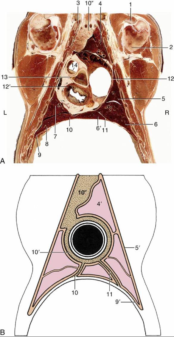
FIG. 27.5 Dorsal section of the bovine thorax directly ventral to the shoulder joint. (A) Actual. L, Left side; R, right side. (B) Schematized to show the asymmetry of the cranial and caudal parts of the mediastinum (stippled). 1, Biceps tendon; 2, humerus; 3, first rib; 4, cranial lobe of right lung; 4', pulmonary pleura; 5, middle lobe of right lung; 5', costal pleura; 6 and 6', caudal and accessory lobes of right lung, respectively; 7, caudal part of cranial lobe of left lung; 8, caudal lobe of left lung; 9, diaphragm; 9', diaphragmatic pleura;
10, 10', and 10", caudal, middle, and cranial mediastinum, respectively, the last occupied by the thymus; 11, plica venae cavae; 12 and 12', right and left atrioventricular valves, respectively; 13, left coronary
artery arising from aortic valve; 14, pulmonary valve.
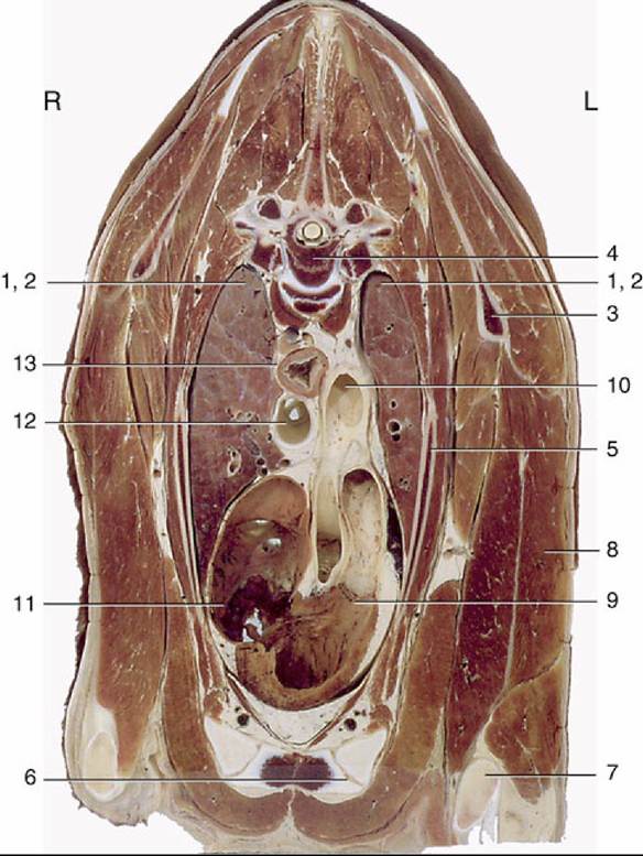
FIG. 27.6 Transverse section of the bovine thorax at the level of the fourth thoracic vertebra. Note the asymmetry of the lungs. 1 and 2, Cranial lobes of right and left lungs; 3, scapula; 4, fourth thoracic vertebra; 5, third rib; 6, sternum; 7, olecranon; 8, long head of triceps; 9, pulmonary valve; 10, aortic arch;
11, right atrioventricular valve; 12, trachea; 13, esophagus; L, left side; R, right side.
Pericardiocentesis is most safety performed in the 5th intercostal space of the left side, directly dorsal to the costochondral joints.