THE HINDGUT
The hindgut develops into the descending colon and the rectum, parts supplied by the caudal mesenteric artery in the adult. Initially the gut ends blindly against the cloacal plate.
Except in the horse and ruminants, in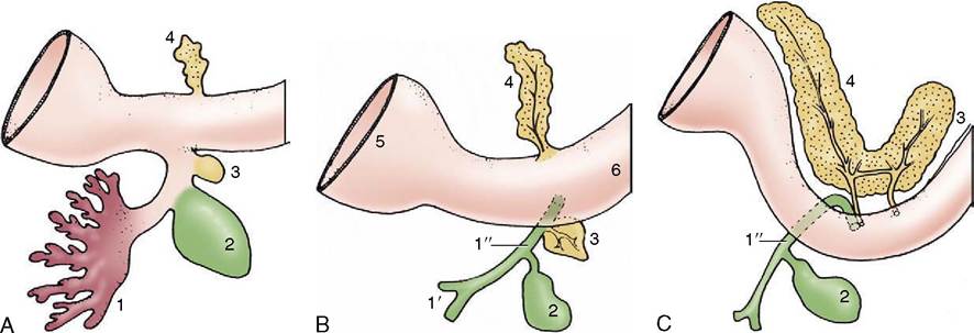
Figure 3-63 Development of the pancreas. A, Early stage. B, A later stage showing separate duct systems in the two primordia. C, The two primordia have fused after the migration of the ventral pancreas. The dorsal pancreas now drains mainly via the ventral duct system. 1, Liver primordium; 1', hepatic ducts; 1", bile duct; 2, gallbladder; 3, ventral primordium of pancreas; 4, dorsal primordium of pancreas; 5, stomach; 6, duodenum.
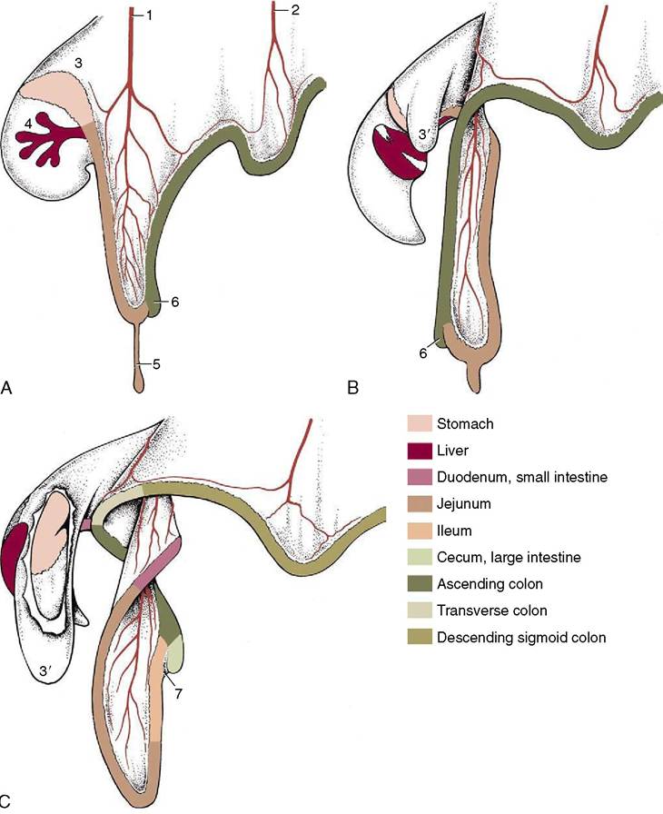
Figure 3-64 Three stages in the growth and rotation of the canine midgut, in left lateral views. 1, Cranial mesenteric artery; 2, caudal mesenteric artery; 3, dorsal mesogastrium; 3', greater omentum, fenestrated in C to expose stomach; 4, ventral meso- gastrium with developing liver; 5, vitelline duct; 6, cecal primordium; 7, ileocecal fold.
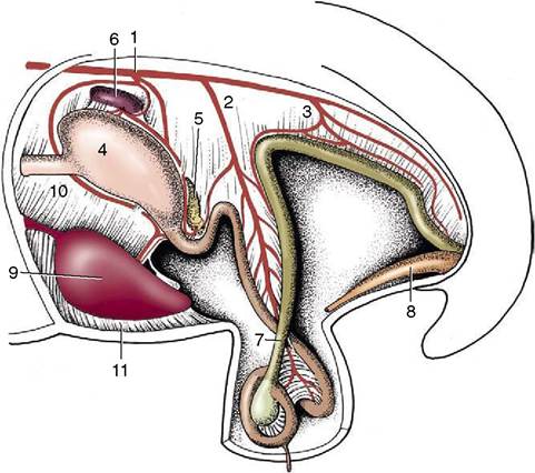
Figure 3-65 Development of the intestinal tract during the rotation process. The midgut loop is herniated into the extraem- bryonic celom. 1, Celiac artery; 2, cranial mesenteric artery; 3, caudal mesenteric artery; 4, stomach; 5, pancreas; 6, spleen; 7, loop of midgut; 8, bladder expansion of the urogenital sinus; 9, liver; 10, lesser omentum; 11, falciform ligament.
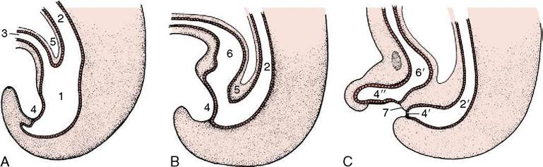
Figure 3-66 Division of the distal part of the hindgut into rectum and urogenital sinus. A, Formation of the allantois and beginning of the caudal extension of the urorectal septum (5).
B, The urorectal septum now approaches the cloacal membrane. C, Complete division of urogenital sinus and anorectal canal. 1, Cloaca; 2, hindgut; 2', anorectal canal; 3, allantois; 4, cloacal membrane; 4,, anal membrane; 4", urogenital membrane; 5, urorectal septum; 6, primitive urogenital sinus; 6,, urogenital sinus; 7, tissue bridge ventral to future anus.which the descending colon shows a secondary increase in length, significant changes affect only the terminal part of the hindgut. A bud, the allantois, grows from its ventral aspect toward and through the umbilical opening in the abdominal wall; once outside the embryo it enlarges to form the capacious allantoic sac (Figure 5-66). A wedge of tissue (urorectal septum) enlarging in the angle between the gut and this diverticulum thrusts toward the cloacal membrane (Figure 3-66). When it meets this, it divides the gut into two separate tubes: the dorsal one is continuous with the descending colon, and the ventral one is continuous with the allantois and destined to form the lower urogenital tract. Meanwhile, proliferation of mesoderm beneath the ectoderm around the proctodeum has deepened the pit; when the dorsal part (anal membrane) of the cloacal membrane breaks down, this deepening is added to the gut, which provides it with the anal canal that leads to the exterior.