THE HINDLIMB
Palpable skeletal features of this limb include the coxal tuber (a slight enlargement at the ventral end of the iliac crest) and the ischial tuber (lateral to the vulva in the female); the greater trochanter of the femur (less readily palpated as it is more deeply placed); the patella, single patellar ligament, the crest and extensor groove of the tibia, and the collateral ligaments, at the stifle; the entire medial surface of the tibia in the leg; and the calcaneus and calcaneal tendon and the medial and lateral malleoli (and adjacent part of the fibula) at the hock (Figure 32-1).
The use of the hamstring muscles for intramus-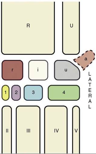
Figure 36-1 The bones of the carpal skeleton in the pig. Roman numerals identify the metacarpal bones, Arabic numerals, the distal carpal bones. R, Radius; U, ulna; a, accessory carpal bone; i, intermediate carpal bone; r, radial carpal bone; u, ulnar carpal bone.
cular injection is contraindicated because of the risks of an adverse effect on the quality of the ham and because of injury to the sciatic nerve.
It is usually impossible to find the subiliac lymph nodes (Figure 36-5/5) located at the cranial border of the thigh, but the popliteal nodes (Figure 36-5/7) may often be palpated, depending on how deeply they lie within the popliteal fossa.