» The Hindlimb
Palpable skeletal features of this limb include the coxal tuber (a slight enlargement at the ventral end of the iliac crest) and the ischial tuber (lateral to the vulva in the female); the greater trochanter of the femur (less readily palpated as it is more deeply placed); the patella, the single patellar ligament, the crest and extensor groove of the tibia, and the collateral ligaments, at the stifle; the entire medial surface of the tibia in the leg; and the calcaneus and calcaneal tendon and the medial and lateral malleoli (and adjacent part of the fibula) at the hock (see Fig.
32.1). The use of the hamstring muscles for intramuscular injection is contraindicated because of the risks of an adverse effect on the quality of the ham and because of injury to the sciatic nerve.It is usually impossible to find the subiliac lymph nodes (Fig. 36.5/5), located at the cranial border of the thigh, but the popliteal nodes (Fig. 36.5/7) may often be palpated, depending on how deeply they lie within the popliteal fossa.
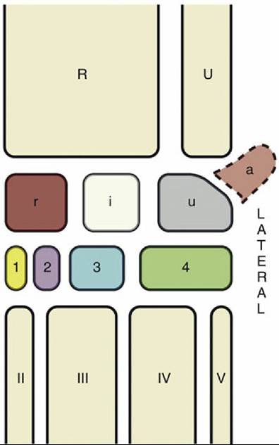
FIG. 36.1 The bones of the carpal skeleton in the pig. Roman numerals identify the metacarpal bones, Arabic numerals the distal carpal bones. a, Accessory carpal bone; i, intermediate carpal bone; r, radial carpal bone; R, radius; U, ulna; u, ulnar carpal bone.
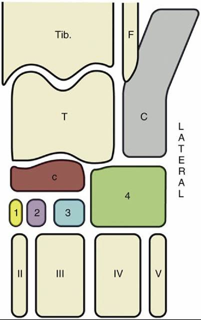
FIG. 36.2 The bones of the tarsal skeleton in the pig, schematic. Roman numerals identify the metatarsal bones, Arabic numerals the distal tarsal bones. C, Calcaneus; c, central tarsal bone; F, fibula; T, talus; Tib., tibia.
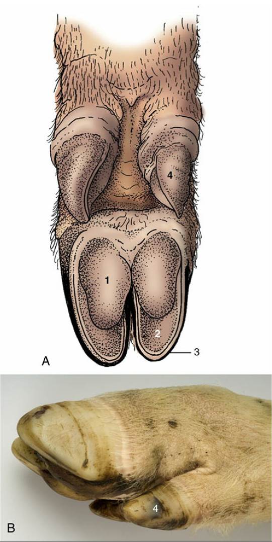
FIG. 36.3 (A) Palmar surface of the foot of a pig.
1, Bulb (digital pad) of hoof; 2, sole of hoof; 3, wall of hoof; 4, hoof of accessory digit. (B) Lateral view of foot of a pig.The Hip Joint
Because of its deep situation, the available landmarks are at some distance from this joint. Depending on the size of the pig, the greater trochanter is located from 2 to 4 cm ventral to the line joining the coxal with the lateral part of the ischial tuber. The needle is inserted at the same distance cranial to the trochanter and passed, at right angles to the skin, through the gluteal muscles to enter the dorsal part of the joint. The greater resistance offered by the fibrous tissue of the deep gluteal muscle and the joint capsule warns that the cavity is close.
The Stifle Joint
The three compartments of this joint communicate, which allows a single injection to reach all parts (see the dog in Fig. 2.63 for the general idea). The puncture is made lateral to the patellar ligament, about one-third of the distance down from the patella to the tibial tuberosity.
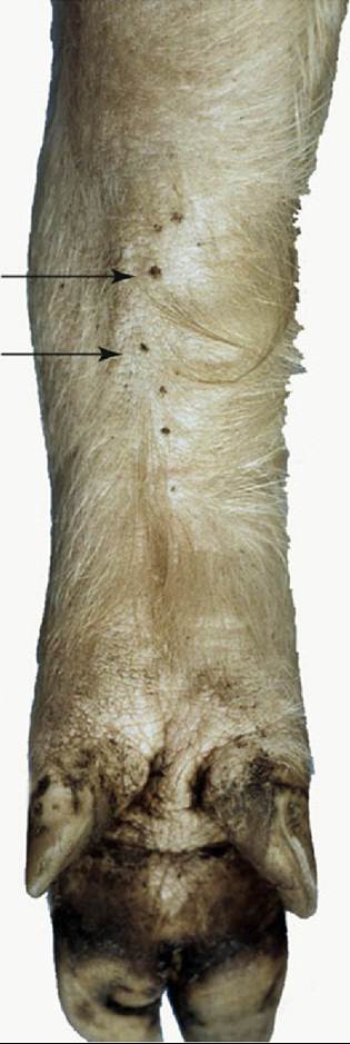
FIG. 36.4 Carpal glands (arrows) of a pig, palmar view.
The Hock Joint
The tarsocrural and proximal intertarsal joints, the only joint compartments at the hock accessible for injection, do not communicate. Two sites are available for injection of the tarsocrural joint, both on the lateral side: one is dorsal and the other is plantar to the collateral ligament. The proximal intertarsal joint is entered from the medial side, plantar to the collateral ligament. There are two independent joint spaces at the tarsometatarsal level: one is proximal to metatarsals II and III, and the other is proximal to metatarsals IV and V. The first of these communicates with the distal intertarsal joint (Fig. 36.2).
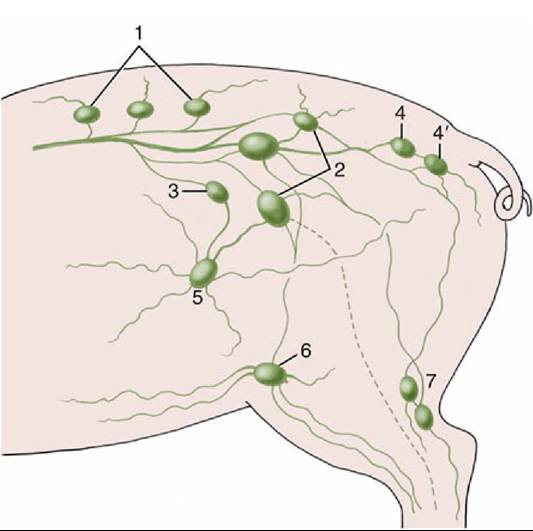
FIG. 36.5 Lymph flow of the hindlimb, lateral view. 1, Lumbar aortic nodes; 2, medial iliac nodes; 3, lateral iliac node; 4, ischial node; 4', gluteal nodes; 5, subiliac nodes; 6, superficial inguinal nodes; 7, popliteal nodes.
No account will be given of the arteries of the limb. Lymph from superficial structures of the thigh and leg drains to the superficial inguinal and subiliac nodes (Fig. 36.5); that from deeper parts travels in lymphatic vessels that run with the major arteries to reach the medial iliac nodes. Lymph from the distal part of the limb drains to the popliteal nodes. Some efferents from these nodes proceed to the gluteal and ischial nodes on the lateral surface of the sacrosciatic ligament; others join the lymphatics running to the medial iliac nodes.
Comprehension Check
Compare the anatomy of the distal limb of the pig, including nerve supply, with that of the horse.