The Hindlimb of the Ruminant
The angular appearance of the hindquarters of cattle is due in part to the robust formation of the pelvic girdle, much of which is outlined below the skin, and in part to the weak development of the muscles of the croup.
The sacral tuber is palpable to the side of the lumbosacral space even though it fails to reach the height of the sacral crest. (Its occasional elevation above the crest prompts suspicion of sacroiliac dislocation.) This tuber is joined to the much more prominent coxal tuber ("hook bone") by the iliac crest, which is only thinly—and incompletely-covered by the gluteus medius (Figs. 31.1 and 31.2). The triangular ischial tuber ("pin bone") is raised considerably above the pelvic floor and projects largely or wholly above the vulva. Its subcutaneous dorsal angle is joined by the sacrotuberous ligament, which is readily palpable because of lack of muscle cover (Fig. 31.3/1').The line connecting the coxal and ischial tubers reveals the slope of the pelvis. An angle larger than usual is associated with a more upright pelvic inlet. A smaller angle (flattened rump) makes the femur more vertical and may predispose to concussive trauma of the hip joint. The hip joint, s position is deduced from the palpation of the greater trochanter situated lateral and slightly caudal to the femoral head, below the intertuberal line (Fig. 31.3/2). The disturbance of this relationship suggests fracture of the neck or dislocation of the head of the femur. Dislocation may occur in several directions and may be due to the relative weakness or occasional absence of the sole intraarticular ligament (ligamentum capitis). Most commonly the trochanter is displaced dorsocranially to project above the intertuberal line. This joint is nominally a ball-and-socket joint, but the extension of the femoral articular surface onto the semicylindrical neck makes it evident that flexion and extension must be the principal movements.
However, the degree of outward rotation of the thigh that accompanies flexion ensures that the stifle is carried free of the abdomen. The cavity of the joint may be reached if a needle is inserted directly in front of the trochanter and is advanced medially and slightly cranially. The deep location and contractions of the muscle pierced en route make the procedure difficult to accomplish successfully.Disorders of the Hip Joint: The hip joint may suffer from luxation, septic arthritis, or fracture of the head of the femur. Luxations occur more in a craniodorsal compared to a caudoventral direction; the latter will likely lodge the dislocated head of the femur in the obturator foramen.
The most striking features of the regional muscles are the relative weakness of the gluteal group and the absence of vertebral origins of the semitendinosus and semimembranosus. The gluteus superficialis is wholly incorporated within the biceps to form the combination sometimes known as gluteobiceps. The gluteus medius possesses a well-defined deep division (gluteus accessorius) with its own insertion tendon protected by a synovial bursa where it passes lateral to the greater trochanter. The biceps fills the caudolateral part of the thigh and has a wide insertion spread between the fascia lata, patella, lateral patellar ligament, and, via the crural fascia, the tibia and calcaneus. A large bicipital bursa intervenes between the lateral epicondyle of the femur and the part of the insertion proceeding to the patellar ligament. The bursa, which may communicate with the stifle joint cavity, is sometimes the site of a painful inflammation, most often encountered in cattle required to rest on bare concrete. The insertions of the semitendinosus and semimembranosus and the actions of the group follow the usual pattern.
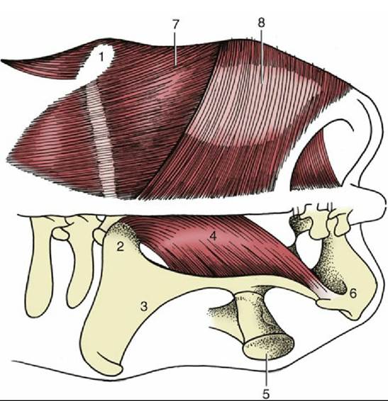
FIG. 31.1 Dorsal view of the bovine croup; the muscles on the left side have been removed.
1, Coxal tuber; 2, sacral tuber; 3, ilium; 4, sacrosciatic ligament; 5, greater trochanter of femur; 6, ischial tuber; 7, gluteus medius; 8, biceps.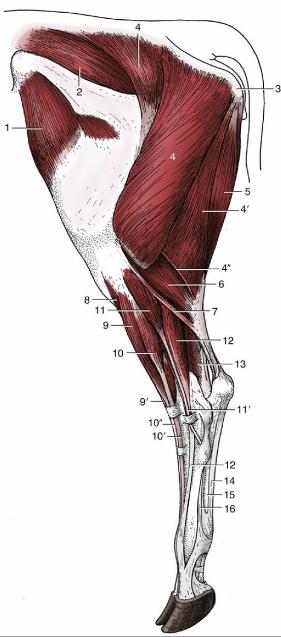
FIG. 31.2 Muscles of the bovine left hindlimb, lateral view. 1, Tensor fasciae latae; 2, gluteus medius; 3, ischial tuber; 4, 4', and 4", biceps, transected at 4"; 5, semitendinosus; 6, lateral head of gastrocnemius; 7, rudimentary soleus; 8, tibialis cranialis; 9 and 9', peroneus tertius; 10, 10', and 10", long digital extensor; 11 and 11', peroneus longus; 12, lateral digital extensor; 13, lateral digital flexor; 14, tendon of superficial digital flexor; 15, combined tendon of deep digital flexors; 16, interosseous.
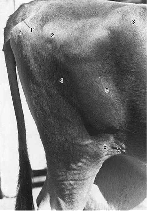
FIG. 31.3 Right bovine thigh. 1, Ischial tuber; 1', Sacrotuberous part of sacrosciatic ligament; 2, greater trochanter of femur; 3, coxal tuber; 4, biceps; 5, lateral vastus; 6, patella; 7, flank fold.
The adductor muscles of the medial thigh, the deep group about the hip joint, and the quadriceps femoris require no special notice. The tensor fasciae latae at the cranial margin of the thigh is a guide to the location of the subiliac lymph node.