The Hypophysis
The hypophysis (pituitary gland; Figs. 8.21 and 8.52), which is attached to the hypothalamus by the infundibulum, has two parts. One, the neurohypophysis (or posterior lobe), is an outgrowth of the brain itself; the other, the adenohypophysis, develops from oral ectoderm (p.
203) and consists of anterior and intermediate lobes. Interspecific differences in the topographic interrelationship of the lobes are not of present concern (see Fig. 6.2).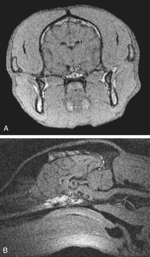
FIG. 8.52 (A) Transverse image at the level of the pituitary fossa and (B) median image of 1-mm-thick T1-weighted gradient-echo magnetic resonance slices of the canine head. 1, Lateral ventricle; 2, basal cistern; 3, pituitary gland; 3', infundibulum; 4, fat in sphenoid bone; 5, third ventricle; 6, interthalamic adhesion; 7, cerebellum; 8, dorsum sellae; 9, pons.
The three lobes produce or store several hormones (p. 204). The posterior lobe hormones (vasopressin and oxytocin) are produced by neurosecretory cells within the supraoptic and paraventricular nuclei of the hypothalamus and are conveyed along the axons for direct release into the neurohypophysial capillary bed (see Fig. 6.3).