The hypophysis or pituitary gland, sometimes called the master gland, produces certain hormones that directly influence the activities of other endocrine glands.
It acts as the relay between the nervous and humoral mechanisms that jointly control certain functions.
The hypophysis is a dark ellipsoidal body measuring about 1 ? 0.75 ? 0.5 cm in the medium-sized dog.
It is suspended below the hypothalamus by a narrow, fragile stalk and is received into a depression (hypophyseal fossa or sella turcica) of the cranial floor that is defined by rostral and caudal crests of bone. A covering of dura directly invests the gland and also roofs the depression, extending from its margins to embrace and confine the hypophyseal stalk from all sides; this arrangement (diaphragma sellae) makes it exceedingly difficult to remove the brain at autopsy with the hypophysis attached.Features of Clinical or Experimental Interest A large venous channel (cavernous sinus) to each side of the hypophysis provides a longitudinal connection between the ophthalmic plexus (and thus the veins of the face) rostrally and the external jugular vein and vertebral venous plexus caudally (p. 300); transverse (intercavernous) sinuses rostral and caudal to the gland complete an encircling venous ring. The internal carotid artery (or the emissary vessel from the rete mirabile that replaces this in the cat, ruminants, and pig [p. 298]) runs through the cavernous sinus to join the arterial circle below the brain. The optic chiasm is directly rostral to the hypophysis (see Fig. 8.22/21 and 24), and laterally, flanking the cavernous sinus, are the cranial nerves that supply the adnexa of the eye (the oculomotor, trochlear, ophthalmic, and abducent nerves).
Pathologic growth or a physiologic increase in the size of the hypophysis, which occurs in pregnancy, may exert pressure on these structures, especially on the optic nerves. Specific features in topography affect both the manner of expansion and the most convenient surgical approach.
This approach is made via the nose and the sphenoidal sinus (within the cranial base, rostroventral to the hypophyseal fossa) in human patients but more directly from below, via mouth, pharynx, and sphenoid, in the dog. A temporal approach has been used in the pig.Although the hypophysis appears to be a solid unitary organ, its parts have very different origins and functions, and it includes certain spaces. One part, the neurohypophysis (posterior lobe), is formed by a downgrowth of the hypothalamus; the stalk that persists as the connection with the brain includes an extension of the third ventricle. The other part, the adenohypophysis (anterior lobe), is formed by an epithelial outgrowth of the roof of the developing mouth. It contains a flattened vestigial space, the hypophyseal cleft. The tissue caudal to the cleft is directly applied to the neurohypophysis and is distinguished as the pars intermedia (intermediate lobe). The topographical relationships of the three "lobes" show some interspecific differences (Fig. 6.2).
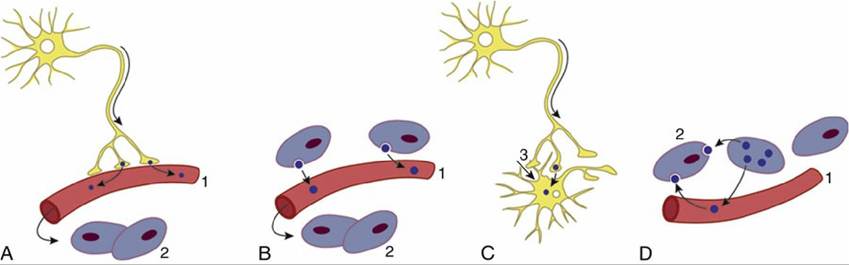
FIG. 6.1 The various ways in which peptides reach their targets: (A) neuroendocrine, (B) endocrine, (C) neurotransmitter, neuromodulator (action on postsynaptic membrane), and (D) paracrine (localized hormone action). 1, Bloodstream; 2, target cell; 3, synapse.
The adenohypophysis produces several hormones commonly designated by acronyms: growth (somatotropic) hormone (STH); gonadotropic hormones—follicle-stimulating (FSH) and luteinizing (LH); adrenocorticotropic hormone (ACTH); thyroid-stimulating hormone (TSH); and prolactin. The intermediate part produces α-melanocyte-stimulating hormone (MSH). The production of all these hormones is controlled by regulating hypophysiotropic hormones and releasing or inhibitory factors such as gonadotropin-releasing hormone (GnRH), somatostatin (SS), growth hormone- releasing hormone (GRH), and corticotropin-releasing hormone (CRH), to name the most important. They are produced by neurosecretory cells in several hypothalamic nuclei, particularly the paraventricular nucleus, preoptic area, arcuate nucleus, and periventricular nucleus.
These hormones are secreted from their axon terminals and are discharged into fenestrated capillaries within the median eminence (see Fig. 8.67/6); these releasing and inhibitory hormones are conveyed to a sinusoidal network within the adenohypophysis (Fig. 6.3).The hormones stored and later released into the circulation by the neurohypophysis include oxytocin and vasopressin. Oxytocin stimulates contraction of the smooth muscle of the uterus and the myoepithelial cells of the udder. Vasopressin stimulates vasoconstriction and promotes fluid reabsorption by the kidneys. These substances are produced by magnocellular neurosecretory neurons within the supraoptic and paraventricular nuclei of the hypothalamus and are conveyed along the axons for direct release via the neurohypophyseal capillary bed into the main circulation.
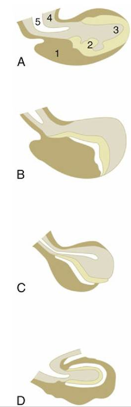
FIG. 6.2 Median sections of the hypophysis of the (A) horse, (B) ox, (C) pig, and (D) dog. The rostral extremity of the gland is to the left. 1, Adenohypophysis; 2, intermediate part; 3, neurohypophysis; 4, hypophyseal stalk; 5, recess of third ventricle.
The neurohypophysis is supplied by small branches from the internal carotid artery (or substitute
vessel) and the arterial circle (of Willis) of the brain. The adenohypophysis is supplied indirectly; rostral hypophyseal arteries, also from the internal carotid, expend themselves within the floor of the hypothalamus, whence the blood is conveyed through the stalk by a portal system of veins. The capillary network of the adenohypophysis subsequently drains into the cavernous sinus.
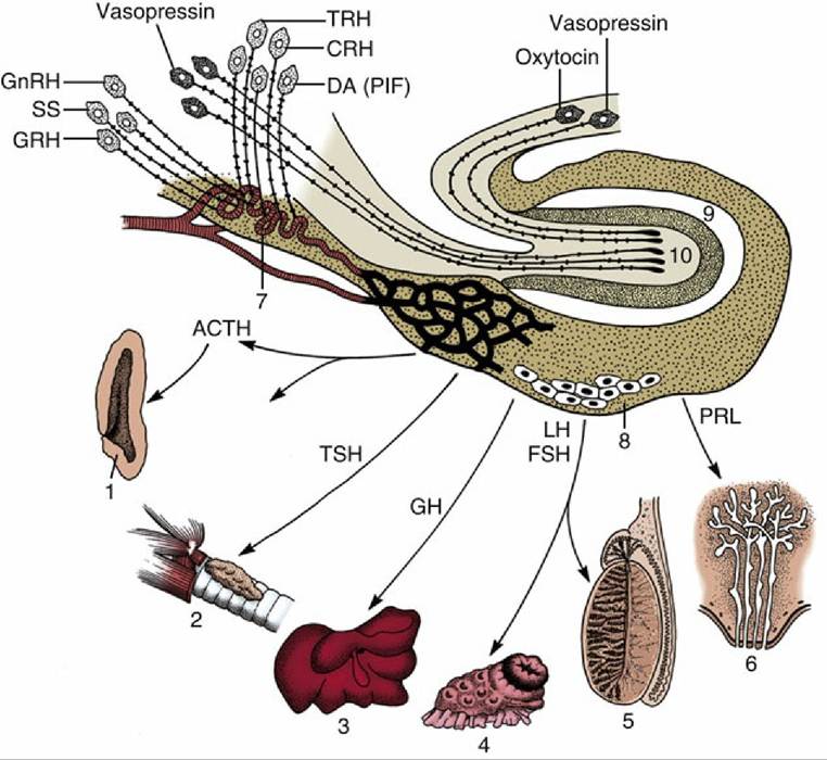
FIG. 6.3 Organization of the brain-pituitary-peripheral organ axis. ACTH, adrenocorticotropic hormone; CRH, corticotropin-releasing hormone; dA, dopamine; FSH, follicle-stimulating hormone; GH, growth hormone; GnRH, gonadotropin-releasing hormone; GRH, growth hormone-releasing hormone; TH, luteinizing hormone; RIF, prolactin-inhibiting factor; PRL, prolactin; SS, somatostatin; TRH, thyrotropinreleasing hormone; TSH, thyroid-stimulating hormone.
1, Adrenal cortex; 2, thyroid; 3, liver; 4, ovary; 5, testis; 6, mammary gland; 7, median eminence; 8, anterior lobe of pituitary; 9, intermediate lobe of pituitary; 10, neural lobe of pituitary.Certain regions of the brain, collectively known as the Circumventricular organs (CVOs), directly receive chemosensory stimulation from bloodborne substances. This occurs as a result of the fenestration of perfusing capillaries, unlike the tight blood-brain barrier elsewhere, which allow exchange of large molecules between the plasma and the extracellular milieu of the CVOs. The proximity of CVOs to the system of ventricles within the brain also suggests a role for the cerebrospinal fluid in the diffusion of the chemical messengers. The neurons within the different regions are of course able to communicate through synaptic connections in the usual way but also allow CVOs to use neurohormonal mechanisms to influence peripheral function. The CVOs comprise the subfornical organ, the pineal gland, the subcommissural organ, the area postrema, the posterior lobe of the pituitary, the median eminence, and the vascular organ of the lamina terminalis (see Fig. 8.67). These organs are broadly concerned with homeostatic and autonomic function (feedback regulation) and induce peripheral effects through secretion of chemicals that are carried into the general circulation by the fenestrated capillaries.
354