THE INTESTINES
The intestines occupy the caudal part of the body cavity, making extensive contact with the gizzard and reproductive organs (Figure 37-16). They consist of duodenum, jejunum, ileum, and colon that open into the cloaca.
In herbivorous birds there also two ceca that arise from the ileocolic junction and accompany the ileum in retrograde fashion (Figure 37-20).The duodenum passes caudally from the right surface of the gizzard. It forms a tight U-shaped loop that returns the duodenojejunal junction to the vicinity of the stomach. Most of the loop lies on the abdominal floor and follows the caudal curvature of the gizzard (Figure 37-16). The pancreas lies between the limbs and empties into the distal end of the ascending duodenum; the bile ducts enter close by (Figure 37-20/4).
The jejunum forms loose coils along the edge of the mesentery and is so thin walled that its content causes
1
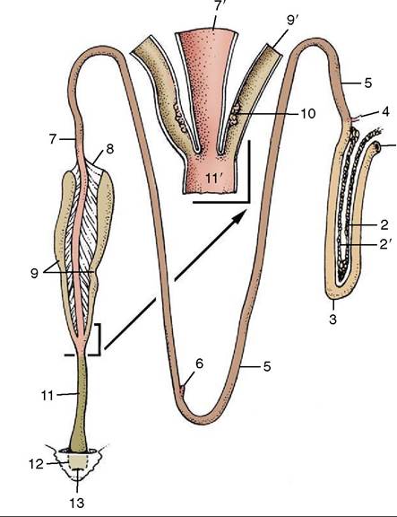
Figure 37-20 Isolated intestinal tract with detail of ileocolic junction. 1, Pylorus; 2, 2', dorsal and ventral lobes of pancreas; 3, duodenal loop; 4, bile and pancreatic ducts entering duodenum; 5, jejunum; 6, vitelline diverticulum; 7, ileum; 7', ileum opened; 8, ileocecal fold; 9, ceca; 9', cecum opened; 10, cecal tonsil; 11, colon; 11, colon opened; 12, cloaca; 13, vent.
it to appear greenish (Figure 37-21, A-B). A small outgrowth (vitelline or Meckel diverticulum; Figure 37-21/8) marks the former connection with the yolk sac. (The yolk sac persists within the body cavity to nourish the hatchling for a few days.) Patches of aggregate lymph nodules are present. In the duck and goose, the jejunum is arranged in several U-shaped loops; in the pigeon, it forms a cone-shaped mass with outer centripetal and inner centrifugal turns. In insect- and fruiteating birds the jejunum is very short and wide.
The ileum continues from the jejunum without demarcation. It is variably described as beginning at the vitelline diverticulum or opposite the apices of the ceca (see Figure 37-20).
The large intestine comprises the ceca and the colon (Figure 37-20/9,11). The ceca, relatively long in the chicken and the turkey, arise at the ileocolic junction and pursue retrograde courses beside the ileum to which they are attached by ileocecal folds. They pass cranially at first, then double back so that their blind ends usually lie near the cloaca (Figure 37-16/13). The proximal
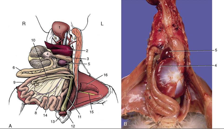
Figure 37-21 A, Gastrointestinal tract after reflection of liver, stomach, and small intestine Craniodextrally, ventral view. B, Detail of stomach and duodenum loop with pancreas within the loop. 1, Crop; 2, left lobe of liver; 3, proventriculus with vagus on dorsal surface; 4, cranial blind sac on right side of reflected gizzard; 5, spleen; 6, duodenal loop enclosing pancreas; 7, jejunum; 8, vitelline diverticulum; 9, ileum; 10, ceca; 11, colon; 12, cloaca; 13, vent; 14, cranial mesenteric vessels and intestinal nerve in mesentery; 15, sciatic nerve and ischial artery; 16, gracilis and adductor.
segment of each has a heavy muscle coat (cecal sphincter) and contains much lymphoid tissue (the so-called cecal tonsil; Figure 37-20/10). The thin-walled middle part appears greenish because of its content. The blind end is thicker walled and bulbous. Bacterial breakdown of cellulose occurs in the ceca. Passerine birds and pigeons have very short lymphoid ceca; psittacines and some carnivorous birds have none.
The (colo)rectum is about 10 cm long in chickens and ends by a slight enlargement at the cloaca. The colorec- tum is no thicker than the small intestine and reabsorbs water and electrolytes by antiperistaltic movements. Urine is moved from the cloaca into the colorectum by antiperistalsis.