THE KIDNEYS AND URETERS
The kidneys are brown and elongated (Figure 37-29 and Figure 37-30). They fill the recesses in the ventral surfaces of the hip bones and lie against the synsacrum, reaching almost to its caudal limit; cranially, they are in contact with the lungs.
The abdominal air sacs that lie against their ventral surfaces extend diverticula that penetrate the dorsal renal surfaces. Several vessels and nerves pass through the kidneys, which makes it impossible to remove them uninjured. Birds suffering from renal gout (not uncommon in commercial flocks) or tumors (common in budgerigars) may therefore have lameness as the presenting sign.Each kidney is arbitrarily divided into cranial, middle, and caudal divisions by the external iliac and ischial arteries (Figure 37-29/12,18), branches of the abdominal aorta. In some species, but not the chicken, the right and left caudal divisions are fused.
The cortex and medulla are not clearly demarcated, and there is no renal pelvis. The ureter (Figure 37-29/20) arises in the cranial division by the confluence of several primary branches and passes over the medio- ventral surface of the kidney, receiving further branches from the middle and caudal divisions in its passage. The ureter then continues caudally alongside the genital duct to end in the dorsal wall of the urodeum (see later). It obtains a whitish tinge from the concentrated urine within it. Neither bladder nor urethra is present.
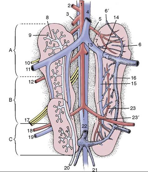
Figure 37-29 Ventral view of the kidneys and vessels and nerves in their vicinity, schematic. The right kidney shows the branches of the ureter; the left, the renal vessels. Cranial (A), middle (B), and caudal (C) divisions of kidney. 1, Aorta; 2, celiac a.; 3, cranial mesenteric a.; 4, caudal vena cava; 5, cranial renal a.; 6, cranial renal portal v.; 6', anastomosis with vertebral venous sinus; 7, cranial renal v.; 8, primary branch of ureter; 9, secondary branch of ureter; 10, femoral n.; 11, external iliac v.; 12, external iliac a.; 13, common iliac v.; 14, portal valve; 15, caudal renal v.; 16, caudal renal portal v.; 17, sciatic n.; 18, ischial a.; 19, ischial v.; 20, ureter; 21, internal iliac v.; 22, caudal mesenteric v.; 23, 23', middle and caudal renal aa.
Each branch of the ureter (Figure 37-29/8) results from the confluence of several secondary branches that receive urine from a small group (five or six) of coneshaped renal lobules, each 1 to 2 mm in diameter. Those near the surface bulge slightly, providing a visible pattern. Each lobule contains nephrons of two types: medullary nephrons resembling the mammalian type (with the loop of Henle) and cortical nephrons resembling the reptilian type together with the vascular networks responsible for extracting urine from the blood. The collecting tubules lie in the periphery of the cone and become confluent at the apex.
The Blood Vessels of the Kidneys
The kidney is supplied by three renal arteries, one for each division (Figure 37-29). The cranial artery arises
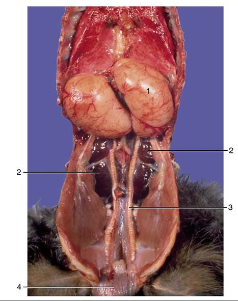
Figure 37-30 Ventral view of the male reproductive organs.
1, Testis; 2, kidney; 3, deferent duct; 4, cloaca.
from the aorta, while the others arise from the ischial artery; together they form the interlobular arteries. Intralobular arteries branch from the interlobular arteries and give rise in turn to two or more afferent arterioles that supply the renal corpuscles (i.e., glomeruli and tubules). However, it is not uncommon for interlobular arteries to give rise to afferent arterioles directly, especially to those supplying close by glomeruli. The smaller veins are satellite to the arteries, but the several renal veins (Figure 37-29/7,15) leaving the organ join the common iliac vein (Figure 37-29/13) and, via this, the caudal vena cava. Superimposed on this is a portal system comprising cranial and caudal renal portal veins (Figure 37-29/6,16). These receive blood from caudal parts of the body (through the external iliac vein) and channel it to the intralobular capillary beds that also receive arterial blood from the renal arteries. Thus, blood that has already passed one capillary bed (in the hindlimb or the pelvis) passes through a second bed within the kidneys. A portal valve (Figure 37-29/14) (situated peripheral to the union of the external iliac and caudal renal veins to form the caudal iliac vein) regulates the flow of blood from the external iliac vein to the kidney; when it is narrowed, more blood enters the kidneys, although some always escapes via connections with the vertebral sinuses and caudal mesenteric vein (Figure 37-29/6',22) at the cranial and caudal ends of the system. Most blood in the caudal mesenteric vein passes through the right hepatic portal vein and the liver before arriving at the heart. (Because of this, it has been suggested that antibiotics should not be injected into the muscles of the hindlimb, as some of the drug would then be excreted by the kidney before reaching the heart for general distribution.)