The Kidneys and Adrenals
The kidneys lie against the diaphragm and psoas muscles dorsally, each enclosed within a capsule of fat. The right kidney lies ventral to the last two or three ribs and first lumbar transverse process; the left one lies ventral to the last rib and first two or three processes and is thus about half a kidney length caudal to the level of its fellow (Fig.
21.19/10 and 11). Each kidney weighs about 700 g. The right one is shaped like the heart on a playing card, but the left one has a more conventional form. Both are dorsoventrally flattened.The cranial pole of the right kidney fits into the renal impression of the liver; caudal to this it is ventrally attached to the pancreas and the base of the cecum (Fig. 21.15/5 and 2). The duodenum winds around the lateral margin and adjoining part of the ventral surface, which is the only region sometimes covered with peritoneum. The short medial border is indented by the hilus and is related to the caudal vena cava and the right adrenal gland (Fig. 21.22).
The ventral surface of the left kidney has a more complete covering of peritoneum and is related to coils of small colon and small intestine, generally including the duodenojejunal junction. Cranioventrally it lies against the spleen and may make contact with a distended stomach (see Fig. 21.10). The medial border is related to the aorta and the left adrenal gland (see Fig. 21.22).
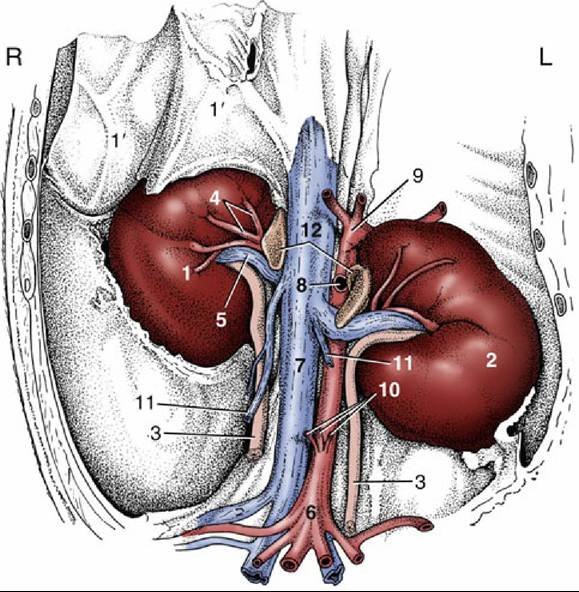
FIG. 21.22 Kidneys and adrenal glands in situ, ventral view. 1, Right kidney; 1', liver; 2, left kidney; 3, ureter; 4, renal artery; 5, renal vein; 6, aorta; 7, caudal vena cava; 8, cranial mesenteric artery; 9, celiac artery; 10, caudal mesenteric and testicular arteries; 11, testicular veins; 12, adrenal glands; L, left; R, right.
The kidneys are of a modified unipyramidal type; the numerous constituent pyramids are completely fused, and their former boundaries are revealed only by the arrangement of the interlobar arteries.
A clearer indication of the lobation, with some external fissuration, is common in the foal. The structure is best revealed in section (Fig. 21.23). The strong external fibrous capsule can normally be easily stripped away, except within the renal sinus, where it merges with the adventitia of the structures entering and leaving. The division of the parenchyma between cortex and medulla is indicated by a color change and by the sectioned arcuate arteries. The cortex is brownish red and granular. The peripheral part of the medulla is dark red, the inner part pale; both show radial striations. The apices of the fused medullary pyramids form a common renal crest that projects into the pelvis. This has a curious form consisting of a central expansion (Fig. 21.23/4) at the origin of the ureter and two terminal recesses toward the poles (Fig. 21.23/5); most papillary ducts open into the recesses. The pelvic mucosa produces a mucous secretion, and as a result the unfiltered urine normally contains some protein (physiologic albuminuria).The renal vessels are short and wide. The artery often splits before reaching the hilus, and a number of branches may enter the ventral surface independently (Fig. 21.23/8).
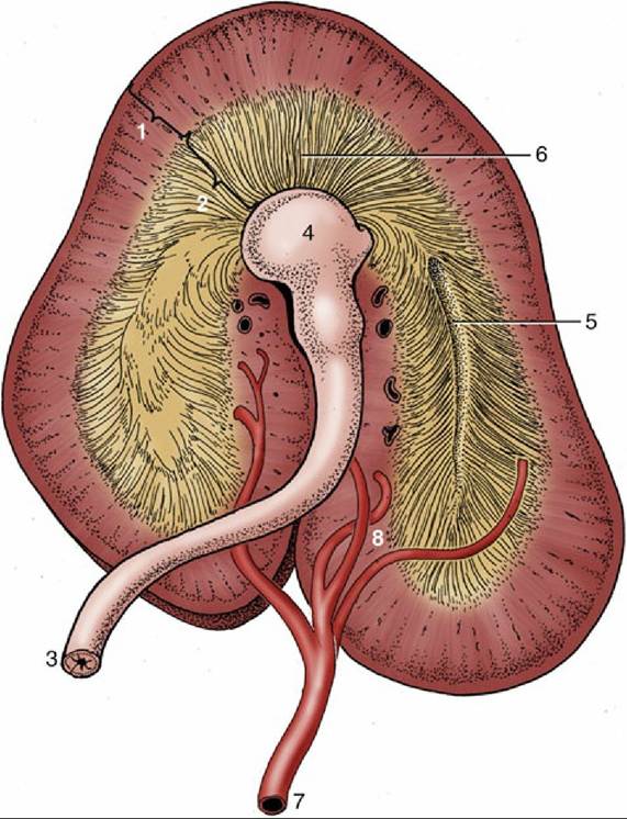
FIG. 21.23 Dorsal section through a kidney (semi-schematic). 1, Renal cortex; 2, renal medulla; 3, ureter; 4, pelvis; 5, terminal recess; 6, papillary ducts; 7, renal artery; 8, interlobar arteries.
For biopsy or complete removal (nephrectomy), the kidneys may be accessed through the 15th or 16th intercostal space or via resection of the 16th or 17th rib.
The ureters are wide at their origins but soon reduce to narrow, more uniform calibers. They bend caudally on emerging from the renal sinus and thereafter pursue a tortuous course over the roof of the abdomen to reach the pelvis. Here they follow the lateral parts of the broad ligaments (genital fold in the male) before inclining medially to pierce the bladder wall close to its neck.
The elongated and irregular adrenal glands lie against the cranial parts of the medial borders of the corresponding kidneys (Fig. 21.22/12). Each consists of an outer bright yellow cortex and an inner brownish red medulla. The glands are relatively large in juvenile animals.
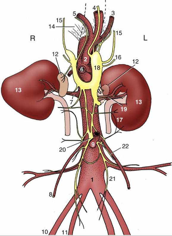
FIG. 21.24 Schema of the abdominal autonomic nerves and branches of the abdominal aorta, ventral view. 1, Aorta; 2, celiac artery (a.); 3, splenic a.; 4, left gastric a.; 5, hepatic a.; 6, cranial mesenteric a.; 7, renal a.; 8, testicular (ovarian) a.; 9, caudal mesenteric a.; 10, external iliac a.; 11, internal iliac a.; 12, adrenal glands; 13, kidneys; 14, crus of diaphragm; 15, major splanchnic nerves; 16, minor splanchnic nerves; 17, lumbar splanchnic nerves; 18, combined celiac and cranial mesenteric ganglia; 19, renal plexus; 20, caudal mesenteric ganglion; 21, hypogastric nerve; 22, testicular (ovarian) plexus; L, left; R, right.