The Liver and Pancreas
The avian liver is dark brown (except in the first 2 weeks after hatching, when it obtains a yellow color from yolk pigments, which continue to be absorbed from the intestine before the yolk sac finally regresses).
It consists of right and left lobes, connected cranially by a bridge dorsal to the heart (Fig. 37.16). Because there is no diaphragm, the lobes of the liver embrace the caudal portion of the heart. The larger right lobe carries the gallbladder on its visceral surface and is perforated by the caudal vena cava; the left lobe is divided (Fig. 37.24). The convex parietal surface lies against the sternal ribs and sternum and is exposed when the breast muscles and sternum are removed in postmortem examination. The liver is covered by a peritoneal sac (cava peritonaei hepatis) that can contain much fat, and fills with transudate in certain diseases. The concave visceral surface makes contact with the spleen, proventriculus, gizzard, duodenum, jejunum, and ovary (or right testis). Two bile ducts, one from each lobe, enter the distal end of the duodenum close to the pancreatic ducts; only the duct from the right lobe is connected to the gallbladder. Pigeons, most parrots, budgerigars, and Struthioniformes lack a gallbladder. Except near the hilus, the hepatic lobules are indistinct because of the lack of perilobular connective tissue.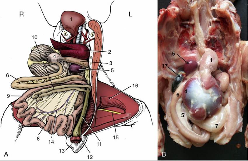
FIG. 37.21 (A) Gastrointestinal tract after reflection of liver, stomach, and small intestine Craniodextrally, ventral view. L, Left; R, right. (B) Detail of stomach and duodenum loop with pancreas within the loop. 1, Crop; 2, left lobe of liver; 3, proventriculus with vagus on dorsal surface; 4, cranial blind sac on right side of reflected gizzard; 5, spleen; 5', Pancreas; 6, duodenal loop enclosing pancreas; 7, jejunum; 8, vitelline diverticulum; 9, ileum; 10, ceca; 11, colon; 12, cloaca; 13, vent; 14, cranial mesenteric vessels and intestinal nerve in mesentery; 15, sciatic nerve and ischial artery; 16, gracilis and adductor; 17, Gall bladder.
The elongated pancreas lies between the limbs of the duodenal loop (Fig. 37.20/2 and 2'). It consists of dorsal and ventral lobes distally connected. Two or three ducts convey pancreatic juice into the distal end of the duodenum.