» The Lymphatic Structures of the Head and Neck
The most important lymph nodes of the head were mentioned in their topographic contexts; other smaller nodes that are usually found medial to the ramus of the mandible are of slight practical concern.
The parotid node (Fig. 25.2/13) receives lymph from the skin covering most of the head, especially the more dorsal areas. It also collects from the upper jaw, temporomandibular joint, masticatory muscles, nasal cavity, hard palate, orbit, and the region about the external ear. The efferent vessels pass to the lateral retropharyngeal node (Table 25.4).
The territory of the mandibular node (Fig. 25.2/20) overlaps those of the parotid and medial retropharyngeal nodes. The chief afferent vessels come from the skin and underlying structures of the ventral part of the head and from the rostral part of the mouth, including the apex of the tongue. The efferent vessels pass to the lateral retropharyngeal node.
The large medial retropharyngeal node lies embedded in fat between the pharynx and the muscles below the cranial base (Figs. 25.9/18 and 25.20/12). It collects lymph from most of the deeper structures of the head, including the nasal and oral cavities, pharynx, larynx, cranium, and jaw muscles, and from the ventral part of the upper end of the neck. The efferent vessels once again drain into the lateral retropharyngeal node, which is the collecting center for the entire head (Fig. 25.26/4). This lateral node, which is placed below the atlantal wing (Fig. 25.2/14), also acts as a primary center for additional lymph vessels draining deeper structures of the head. It channels its outflow into a single large vessel, the tracheal duct, that runs down the neck within the fascia covering the lateral aspect of the trachea. The duct ends by joining the thoracic duct or by opening into one or another vein at the thoracic inlet; most usually the left tracheal duct opens into the thoracic duct while the right one drains directly into a major tributary of the cranial vena cava (Fig.
25.26/9).» TABLE 25.4
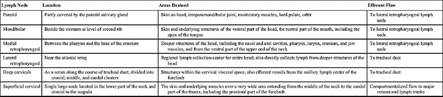
Lymph Nodes of the Head and Neck
A series of small deep cervical lymph nodes is spread along the course of each tracheal duct. These are supposedly divided into cranial, middle, and caudal clusters and receive lymph from the structures within the cervical visceral space. They transmit this lymph to the tracheal duct, sometimes directly and sometimes after serial passage through several nodes within the group. Usually one or more of the most caudal of these nodes receive the efferent vessels of the axillary lymph center of the forelimb, as well as smaller trunks coming directly from the brisket.
A single, much larger node lies in the lower part of the neck in front of the scapula. This is the superficial cervical (prescapular) node (Fig. 25.26/6), which rests on the deep muscles over the cervical vertebrae; it is easily palpated, though covered by the omotransversarius. It collects from the skin and underlying muscles over a very wide area extending from the middle of the neck to the caudal part of the thorax, including the proximal part of the forelimb. The flow through the node is compartmentalized; particular portions of the node are related to different parts of the drainage field. The large efferent vessels open variously into the major lymph and venous trunks in the vicinity.
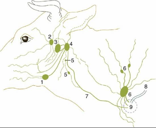
FIG. 25.26 The lymph drainage of the head and neck. 1, Mandibular lymph node; 2, parotid lymph node;
3, medial retropharyngeal lymph node; 4, lateral retropharyngeal lymph node; 5, deep cervical lymph nodes; 6, superficial cervical lymph nodes; 7, tracheal duct; 8, thoracic duct; 9, area within which lymphatic vessels enter veins.
Any of the major nodes may be duplicated.
Comprehension Check
Examine the teeth of the cadavers in the laboratory, and estimate the age of the animals.
Sketch the location of various lymph nodes in the head and neck of cattle, and show the path of lymph drainage.
Use bovine skull, intact and sectioned in various planes, to understand the location and extent of various paranasal sinuses.