The Male Reproductive Organs
These consist of paired testes, epididymides, and deferent ducts and a single phallus that in some species, including chickens, ducks, and ostriches, is the copulatory organ. The testes remain at their sites of origin; spermatic cord, tunica vaginalis, and scrotum are therefore lacking.
Neither accessory reproductive glands nor a urethra exists.The Testis
The bean-shaped testes are relatively large (about 5 cm long) and white during the breeding season (Fig. 37.30/1); however, they shrink to about half that size and become yellowish during the quiescent period (during molt). In some birds, especially Passeriformes, the difference in size is even more dramatic. Attached by short mesorchia, the testes are placed symmetrically against the cranial ends of the kidneys, just caudal to the adrenals, related ventrally to the abdominal sacs, proventriculus, liver, and intestines (Fig. 37.31/3). Removal of the testes (caponizing) to promote fattening may be performed through an incision near the last rib. Before the advent of simpler and safer tests to determine the sex of a bird of a monomorphic species, sexing, at least in larger cage birds, could be performed by introducing an endoscope through a small incision.
The serosa covers a thin tunica albuginea from which a scanty stroma is derived; no mediastinum testis exists. The seminiferous tubules pass to the dorsomedial surface, where they open into the rete testis. The epididymis is not divided into head, body, and tail and appears as a slight bulge on the testis. It is formed by tightly packed efferent ductules that join to form the epididymal duct that transports the spermatozoa to the deferent duct (Figs. 37.30/3 and 37.31/7). The tightly coiled deferent duct arises from the caudal end of the epididymis and accompanies the ureter to the cloaca, where it opens on a low papilla on the lateral wall of the urodeum (Fig.
37.22). The duct shows a slight terminal enlargement (receptacle). During the reproductive period the duct, packed with spermatozoa, appears white. The ejaculate of the cockerel is generally not quite 1 mL. The seminal fluid is elaborated in the testes and by the epithelial cells lining the extratesticular ducts.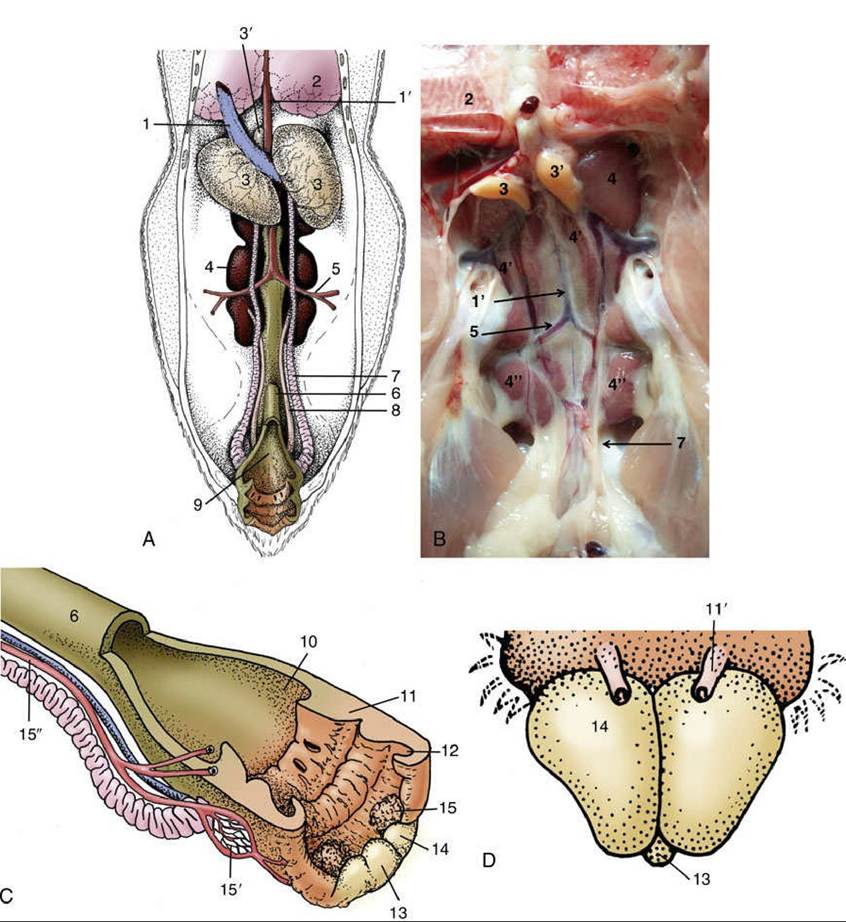
FIG. 37.31 (A) Ventral view of the male reproductive organs (schematic). (B) Ventral view of the male reproductive organs. (C) The floor of the cloaca has been removed and is shown turned over. (D) Caudal
view of the tumescent phallus. 1, Caudal vena cava; 1', aorta; 2, lung; 3, right testis; 3,, left testis; 4, kidney-Cranial lobe; 4', Kidney-Middle lobe; 4", Kidney-Caudal lobe; 5, ischial artery; 6, rectum; 7, deferent duct; 8, ureter; 9, cloaca; 10, coprodeum; 11, urodeum; 11', papilla of right deferent duct; 12, proctodeum;
13, median phallic tubercle; 14, lateral phallic body; 15, lymphatic folds; 15', paracloacal vascular body; 15", pudendal artery.
The Cloaca and Phallus
The coprodeum, the most cranial division of the cloaca, has been described (p. 784). The urodeum (Fig. 37.22/3), caudal to the coprourodeal fold, is indistinctly demarcated from the proctodeum by a shallow, ventrally incomplete uroproctodeal fold (Fig. 37.22/3'). The ureteric orifice is in the dorsolateral wall, above the papilla of the deferent duct. In the female, the slitlike opening of the oviduct (Fig. 37.22/8) occupies a similar position on the left side (see further on). A small patch of vascular tissue (paracloacal vascular body; Fig. 37.31/15') in the lateral wall of the urodeum is thought to supply lymph for the tumescence of the phallus.
The proctodeum—the short, most caudal segment of the cloaca—ends at the vent. A small opening in its dorsal wall leads to the cloacal bursa (bursa of Fabricius; Fig. 37.22/9), an accumulation of lymphatic tissue that is the differentiation site of B lymphocytes (Fig.
37.23). The cloacal bursa is thus an immunologic organ analogous to the thymus (see pp. 797-798). A small (dorsal proctodeal) gland is found caudal to the bursa (Fig. 37.22/9').The vent is a horizontal slit. The ventral lip is of interest because in the male chicken it bears the nonprotrusible phallus, the analogue of the mammalian penis, on its internal surface. The phallus consists of a small median tubercle flanked by a pair of larger lateral phallic bodies (Fig. 37.31/13 and 14). These enlarge in the tumescent state and together form a channel that receives the ejaculate from the deferent ducts (Fig. 37.31C). During insemination, the vent is everted and the phallus is pressed against the cloacal mucosa of the female (cloacal "kiss"). The phallus of the tom turkey is similar. The gander and the drake have a protrusible phallus, several centimeters long and capable of intromission. It is shaped like a thin cone and exhibits a spiral groove that conveys the semen to the tip (Fig. 37.32/8). A protrusible phallus, also seen in ratites, is capable of true intromission into the female cloaca.
Psittacines, passerines, pigeons, and birds of prey all have no phallus. These species copulate by transferring semen from the everted cloaca directly into the female oviduct.
Day-old chicks of both sexes of chickens present a minute genital protuberance at the future location of the phallus. A slight visible difference in form (which is rounded in males and conical in females) is distinguishable by the experienced eye and enables almost all male chicks to be discarded when selecting a laying flock.