THE MENINGES AND CEREBROSPINAL FLUID
1. Visualize the relative location of the meningeal layers to each other and to the skull and brain and to the vertebral canal and spinal cord.
2. What are the arachnoid villi extensions of and what do they extend into?
3.
What is an epidural injection?4. Is cerebrospinal fluid circulated within the epidural space?
5. Which meningeal layer forms the lining of the perivascular spaces and what is its extent?
6. What fluid fills the perivascular space?
7. Visualize the location of the brain ventricles.
8. What structures within the ventricles produce cerebrospinal fluid?
9. Describe the circulation of the cerebrospinal fluid.
0. What could cause the cerebrospinal fluid pressure to increase?
11. What are functions of cerebrospinal fluid?
2. Are blood cells normally not present in cerebrospinal fluid?
Within the bony cranium and vertebral column, the brain and spinal cord have three connective tissue wrappings known as the meninges. In addition, they are cushioned by cerebrospinal fluid, which functions as a shock absorber. Cerebrospinal fluid is formed in the cavities of the brain, the ventricles.
Meninges of the Brain
The meninges are the coverings of the brain and spinal cord. From without inward they are the dura mater, arachnoid, and pia mater, respectively (Figure 4-26). In the skull the outer aspect of the dura mater is intimately fused with the inner periosteum of the calvaria (brain case). Dorsally, between the cerebral hemispheres and between the cerebrum and cerebellum, there is a separation of the outer and inner aspects of the dura mater to form valveless venous sinuses into which drain the veins of the brain and its encasing bone. Venous sinuses are also located beneath the brain, and the paired cavernous sinus plays a big role in the ventral venation of the brain. These blood collection areas are continued as veins that return blood to the heart from the brain.
The only space between the inner aspect of the dura mater and the arachnoid is that which is sufficient for blood vessels. The arachnoid has projections (trabeculae) from its inner aspect to the most intimate covering of the brain, the pia mater. The trabeculae give the appearance of a spider’s web - hence the name arachnoid (after the,class name for spiders, Arachnida). The space between the arachnoid and the pia mater is significant and is known as the subarachnoid space. There are projections from the subarachnoid space into the dura mater sinuses, the microscopic arachnoid villi. Clusters of arachnoid villi, when there are enough to form a macroscopic structure, are called arachnoid granulations. The subarachnoid space contains cerebrospinal fluid and the arachnoid villi allow for the resorption of this fluid back into the blood. The pia mater follows all of the grooves and fissures of the brain surface. It forms a sheath around blood vessels and follows them into the substance of the brain (Figure 4-27). The perivascular spaces thus formed extend as far as the arterioles and venules, but not on to the capillaries. The inner aspects of the brain, therefore, are in communication with cerebrospinal fluid (this might serve a “lymphatic” function because there are no lymph vessels in the brain). The meninges (and cerebrospinal fluid) continue for a short distance on to the cranial and spinal nerves. The vestibular (auditory) branch of the vestibulocochlear nerve (cranial nerve VIII), because of its closeness to the exterior of the body, subjects the meninges to some hazard if the inner ear becomes inflamed.
■ FIGURE 4-26 Cerebral meninges and arachnoid villi. The meninges consist of the dura mater (thickness exaggerated to illustrate dura mater sinus), arachnoid (darkened line), and pia mater. The subarachnoid space (exaggerated as shown) contains cerebrospinal fluid.
The arachnoid villi project into the dura mater sinus (blood sinus) and provide an outlet for cerebrospinal fluid.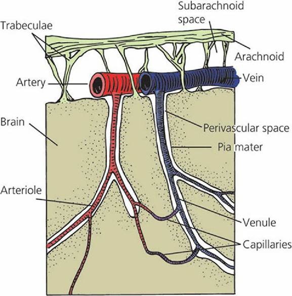
■ FIGURE 4-27 The perivascular space. The space is lined by pia mater that follows blood vessels into the brain substance. The space is filled with cerebrospinal fluid and communicates with the subarachnoid space. It extends only to the level of the capillaries and serves a lymphatic function.
Meninges of the Spinal Cord
The meninges of the spinal cord are continuous with the meninges of the brain. The outer aspect of the dura mater is not fused with the vertebral canal (the hole through the vertebrae within which the spinal cord transcends), and an epidural (outside the dura mater) space exists, which contains fat (Figure 4-28). Sites for entering the epidural space include those between L1-L2 (lumbar vertebrae), lumbosacral, and sacrocaudal for varying purposes and in varying species. The epidural space at the sacrocaudal location is used for the injection of local anesthetics in cattle. Spinal nerve projections are present in this location; when anesthetized, sensory and motor loss occurs in certain areas, which is advantageous for medical or surgical treatment. For example, a prolapsed uterus (uterus that has everted through the vagina) can be replaced in the cow without the straining that would otherwise occur.
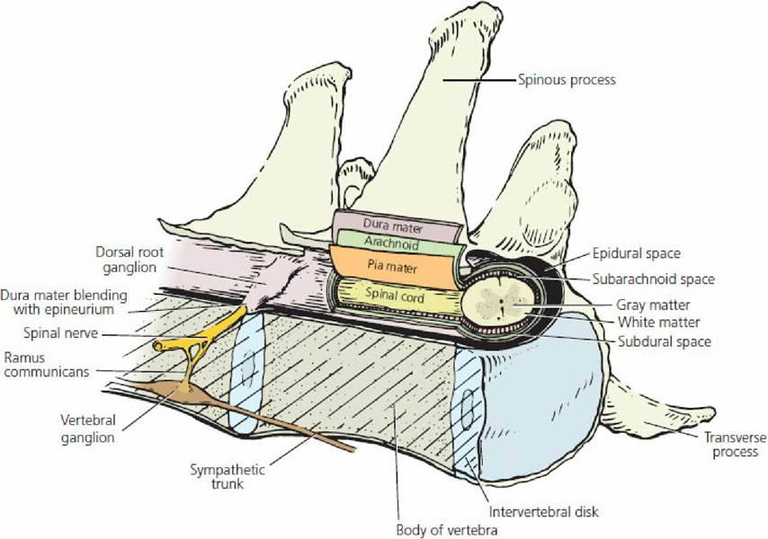
■ FIGURE 4-28 The meninges of the spinal cord. Only half of the vertebra is shown to indicate the extension of the dura mater on to the spinal nerves. Note the presence of an epidural space.
Ventricles of the Brain
The four ventricles of the brain are cavities or hollowed-out spaces within the substance of the brain (Figures 4-29 and Figure 4-30). The lateral ventricles are paired cavities within each right and left cerebral hemisphere.
Each is continuous with the single third ventricle through an interventricular foramen (foramen of Monro). The third ventricle is located within the interbrain and is continuous with the fourth ventricle through the cerebral aqueduct (mesencephalic aqueduct). The fourth ventricle is located beneath the cerebellum and above the medulla oblongata. It communicates in turn with the subarachnoid space by means of paired lateral recesses (see Figure 4-29) and apertures (foramina of Luschka). In addition to the paired foramina of Luschka, primates have a single median aperture (foramen of Magendie). The fourth ventricle is continued caudad as the central canal of the spinal cord. Each of the four ventricles has a structure known as the choroid plexus projecting into it. The choroid plexus is a tuft of capillaries that secretes cerebrospinal fluid. The capillaries belong to the pia mater, but are covered with ependymal cells (a glial cell) that unite with the capillaries to form the choroid plexus.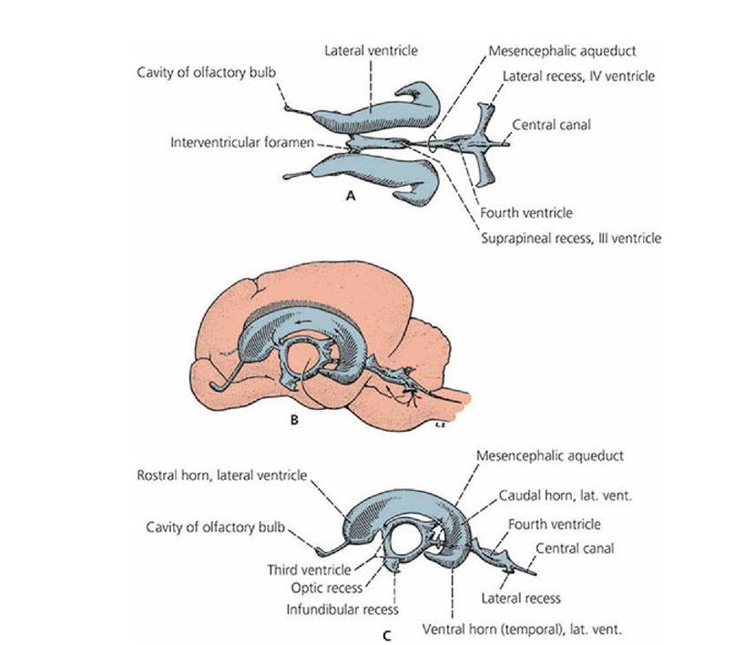
■ FIGURE 4-29 The canine brain ventricles. A. Dorsal view of ventricles without brain substance. B. Lateral view of ventricles, which shows their location within the brain. C. Lateral view of ventricles without brain substance. (From Evans HE, de Lahunta A. A Guide to the Dissection of the Dog. 8th edn. Ames, IA: Wiley-Blackwell, 2017.)
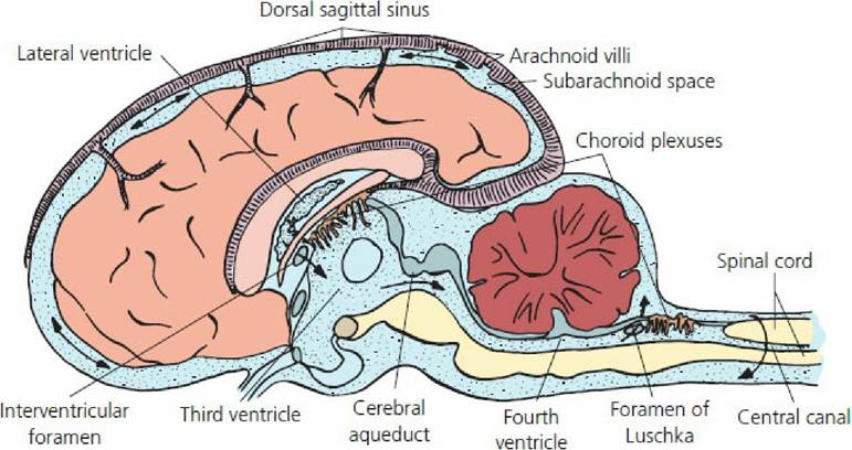
■ FIGURE 4-30 Pathway of cerebrospinal fluid flow from choroid plexuses to the arachnoid villi that protrude into the dural sinuses. The interventricular foramina are openings from each of the two lateral ventricles (one in each cerebral hemisphere). The choroid plexuses produce the cerebrospinal fluid (stippled). The two foramina of Luschka (one shown) provide an exit from the sites of formation to the subarachnoid space of the brain and spinal cord.
Note that cerebrospinal fluid circulates around the spinal cord. Cerebrospinal fluid circulates caudally through the central canal of the spinal cord as a continuation of the fourth ventricle. The central canal may not be patent all the way to caudal levels.Circulation and Function of Cerebrospinal Fluid
Cerebrospinal fluid, formed by the choroid plexuses, flows through the cavities of the lateral and third ventricles, through the cerebral aqueduct and fourth ventricle, and finally through the foramina of Luschka to enter the subarachnoid space of the brain and spinal cord. Cerebrospinal fluid also enters the central canal of the spinal cord from the fourth ventricle. Cerebrospinal fluid then leaves the subarachnoid space of the brain through specialized structures (arachnoid granulations or villi) in which the subarachnoid space invaginates into the cerebral venous sinuses (the dural sinuses) (see Figure 4-26). The relationship of the subarachnoid space with the venous sinuses is such that each villus functions as a valve regulating flow of cerebrospinal fluid into the venous sinus. The backward flow of blood into the subarachnoid space is prevented, but the forward flow of cerebrospinal fluid into the cerebral sinuses is allowed. Therefore, a higher pressure must exist within the subarachnoid space than within the venous system, and removal of cerebrospinal fluid is dependent on the venous pressure being at least 1 mm Hg less than the cerebrospinal fluid pressure. The normal pressure of cerebrospinal fluid ranges from 8 to 12 mm Hg, whereas the pressure within the dural sinuses ranges from 1 to 8 mm Hg.
Cerebrospinal fluid production is fairly constant regardless of pressures within the cranial cavity because it is an active process. Total amounts of cerebrospinal fluid vary with species and size of animal, with dogs producing at a rate around 0.05 mL/min and cats around 0.015 mL/min. In general, mammals produce about three to five times the total volume of their cerebrospinal fluid in a 24-hour period.
If the pathway for flow from the choroid plexuses to the dural venous sinuses is occluded, the cerebrospinal fluid pressure increases and hydrocephalus can result. Cerebrospinal fluid that accompanies the meninges for a short distance on to the cranial and spinal nerves can enter lymphatics at that level and be returned to the blood. This is a particularly important outflow for cerebrospinal fluid that surrounds the spinal cord. In the horse and in sheep, cerebrospinal fluid that enters the central canal has an exit at the caudal extremity via the terminal ventricle. The terminal ventricle then communicates with the subarachnoid space of the spinal cord. A similar arrangement is likely to be present in other animals.The cerebrospinal fluid is thin and watery; it is derived from blood plasma by a secretion process. Except for a few lymphocytes, the normal cellular elements of the blood are absent. In cases of injury or inflammation of the meninges, the number of cellular elements of blood can increase.
The principal function of the cerebrospinal fluid is provision of a watery cushion for the brain and spinal cord. Displacement of the brain is therefore minimized when rapid directional changes occur in the head. The “lymphatic” function (see the previous section) serves the brain and spinal cord for the return of protein that leaks from the capillaries. When blood volume in the brain increases, the volume of cerebrospinal fluid decreases, thereby keeping the volume of cranial contents constant. Determining the pressure of the cerebrospinal fluid can be helpful (e.g., in the neurologic examination of an animal) and is usually about 10 mm Hg.
■