REFLEXES
1. What are the components of a reflex arc?
2. Describe the knee jerk reflex.
3. What is the purpose of the stretch reflex?
4. Why is the stretch reflex considered a postural reflex?
5.
How does the crossed extensor reflex differ from the knee jerk reflex?6. Differentiate between somatic and visceral reflexes.
7. How are visceral reflexes transmitted?
8. List functions of reflex centers located in the: (a) medulla oblongata, (b) cerebellum, (c) hypothalamus, and (d) midbrain.
9. What is meant by muscle tone?
0. What is the basic element of muscle tone?
11. Describe standing, attitudinal, and righting reflexes.
A reflex is defined as an automatic or unconscious response of an effector organ (muscle or gland) to an appropriate stimulus. The components involved for a reflex to occur make up what is known as a reflex arc and consist of: (1) a receptor, (2) an afferent limb, (3) central connections, (4) an efferent limb, and (5) an effector organ.
Spinal Reflex
A reflex can involve parts of the brain and autonomic nervous system, but the simplest reflex is the myotatic (stretch) spinal reflex. An example of a spinal reflex is the knee jerk reflex (Figure 4-25). The reflex is elicited by striking the middle patellar ligament. This ligament, located at the knee, is the tendon of insertion for the quadriceps femoris and transmits its action to extend the tibia. Striking the middle patellar ligament stretches the quadriceps muscle, which in turn stimulates muscle spindles (receptors for muscle sense). An impulse is transmitted by way of the dorsal root of the appropriate spinal nerve to the applicable motor neuron in the ventral horn of the gray matter, and thence to muscle fibers of the quadriceps muscle, causing it to contract. The purpose of the reflex is to oppose stretch of the muscle. Because this reflex involves an intact and functioning spinal cord at a certain level of segmentation, the soundness of the cord at that level can be determined by this reflex action.
Absence of the knee jerk reflex can help to confirm suspicion of damage or injury to the spinal cord or any of the five components of the reflex arc. This reflex is a postural reflex because it aids in maintaining a standing position.
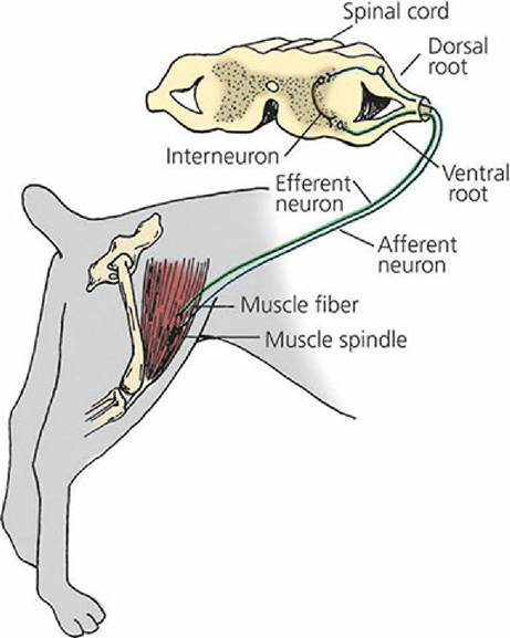
■ FIGURE 4-25 The stretch reflex. Stretch of muscle stimulates the muscle spindle. The impulse travels to the spinal cord by way of an afferent neuron. Transmission of the impulse to an efferent neuron may be direct or by way of an interneuron as shown. Stimulation of an efferent neuron to striated muscle counteracts stretch by causing contraction. Muscle spindles, in addition to being involved in reflexes, also provide sensory input to cerebral and cortical levels as well as playing a role in voluntary control of muscular activity.
Spinal reflexes can also be rather complex, in which the central connections of the reflex extend over several segments and also extend contralaterally as well as ipsilaterally. The crossed extensor response is an example of a complex spinal reflex. This is shown when there is painful stimulation of the skin or subcutaneous tissues and muscle, e.g., touching a hot surface. The response is flexor muscle contraction and inhibition of extensor muscles so that the part that was stimulated is flexed and withdrawn from the stimulus and at the same time there is extension of the opposite limb (assisting withdrawal).
Somatic and Visceral Reflexes
If the effector organs are composed of striated muscle, the reflex is somatic. If the effector organs are either smooth or cardiac muscle, or glands, the reflex is visceral. Visceral reflexes regulate visceral functions and are transmitted by the autonomic nervous system (by visceral afferent fibers and preganglionic and postganglionic efferent fibers of the sympathetic or parasympathetic division).
Reflex Centers
Reflex centers are located throughout the central nervous system.
They are involved with the integration of more complex reflexes. The simplest reflexes are those associated with the spinal cord, and the more complex are carried out through reflex centers in the brain. Some of these centers are located in the pons and medulla oblongata and include reflex centers for the control of heart action, vessel diameter, respiration, swallowing, vomiting, coughing, and sneezing. The cerebellum contains most of the reflex centers associated with locomotion and posture. The hypothalamus is the main integration and regulation center for the autonomic nervous system, e.g., contains reflex centers associated with temperature regulation. The midbrain contains visual and auditory reflexes, which can bring about constriction or dilatation of the pupils and evoke a startle reaction to loud noises.Postural Reflexes and Reactions1

The postural reflexes and reactions aid in maintaining an upright position. Responses that involve the cerebral cortex are more properly called reactions than reflexes. Muscle tonus (tone) is that state of muscle tension that enables an animal to assume and remain in the erect attitude. The stretch reflex, previously described, is the fundamental element of muscle tone. The following are examples of postural reflexes and reactions:
1. Standing reflex - pushing down on the back of a dog causes muscle movements that compensate for and resist the displacement.
2. Attitudinal reflexes - displacement of one part of the body is followed by postural changes in other parts (e.g., lifting the head of a horse is followed by postural changes in the rear quarters so that a new attitude is assumed).
__ ∖,Go to____________________________ to view a related video.
U ■>
3. Righting reflex - dropping an inverted cat is followed by its landing in the upright position.
4. Hopping reaction - pushing a supported dog with three limbs elevated results in a placement correction of the intact leg to act as a rigid pillar.
The spinal cord of domestic animals constitutes a greater proportion of the CNS (brain and spinal cord) than in humans. This reflects the fact that more of the CNS activity in animals is accomplished by reflex than by cerebral activity. There is approximately 10 times more spinal cord activity in dogs than in humans.
■