The Muscle Spindle Stretch Receptor Is an Encapsulated Organ of SpecializedMuscle Fibers with Separate Motor and Sensory Innervations
The muscle spindle is an encapsulated group of about 3 to 12 small, slender, specialized skeletal muscle fibers (Figure 8-2). Because their capsule is spindle shaped, or fusiform, these muscle fibers are called intrafusal muscle fibers.
The muscle fibers that cause physical shortening of the muscle (the majority of muscle fibers in a muscle belly), located outside of the capsule, are called extrafusal muscle fibers. Extrafusal muscle fibers often span the length of the gross muscle from origin to insertion tendon; intrafusal muscle fibers and their capsules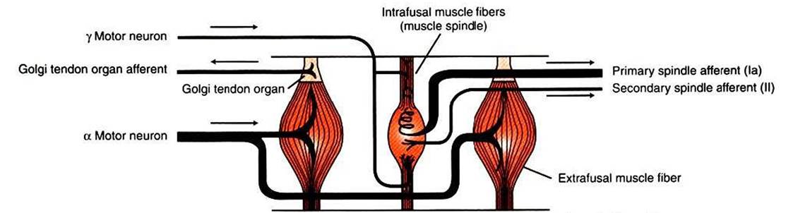
FIGURE 8-1 Skeletal muscles have two important receptors: the muscle spindle and the Golgi tendon organ.The intrafusal muscle fibers (muscle spindle) are arranged in parallel with the e?trafusal muscle fibers; the Golgi tendon organ is in series with the e?trafusal fibers. Arrows indicate direction of action potential flow along respective axons. (Modified from Kandel ER, Schwartz JH: Principles of neural science, ed 2, NewYork, 1985, Elsevier Science Publishing.)
are much shorter (about 4-10 mm long). In addition, the intrafusal muscle fiber endings are attached to the extracellular matrix of, and lie in parallel to, the extrafusal muscle fibers. Therefore, if the muscle is stretched, lengthening the extrafusal muscle fibers, the intrafusal fibers of the muscle spindle are also stretched.
Unlike extrafusal muscle fibers, the contractile elements of intrafusal muscle fibers are restricted to their polar ends, with none in their middle (equatorial) region. Therefore, their polar ends can contract, but their equatorial region cannot. Such contraction does not directly contribute to the shortening of the gross muscle, but it can tighten the region of the intrafusal fiber that lies between the two poles. As discussed later, this can have a dramatic effect on the muscle spindle’s sensitivity for transducing muscle stretch.
Spindle sensory neurons arise from the equatorial region of the intrafusal muscle fibers and carry action potentials from the spindle to the CNS by way of the peripheral nerves. These CNS afferents enter the spinal cord through the dorsal roots (Figure 8-3). The contractile, polar regions of the intrafusal muscle fibers are innervated by motor neurons called gamma (γ) motor neurons. Extrafusal muscle fibers—the muscle fibers that cause the physical shortening of the muscle—are supplied by a different population of motor neurons (those that comprise the motor units) called alpha (α) motor neurons. Although γ motor neurons go to intrafusal muscle fibers and α motor neurons go to the extrafusal muscle fibers, these CNS efferents both have their cell bodies in the ventral horn of the spinal cord, and their axons leave through the ventral roots.