The Muscles of Mastication and the Temporomandibular Joint
The muscles of mastication are well developed. The masseter takes origin along the whole length of the facial crest and zygomatic arch and inserts on the mandible between the vascular notch and condyle (Fig.
18.7/11). It is a multipennate muscle constructed so that the fibers of the superficial strata run caudoventrally, while those more deeply placed are nearly vertical. Its cranial margin produces a very prominent surface contour that serves as a guide to the location of the facial vessels and parotid duct. Its caudodorsal part is overlain by the parotid gland but to a variable depth and extent, which affect the accessibility to palpation of the parotid lymph nodes. Laterally, the masseter is traversed by buccal branches of the facial nerve.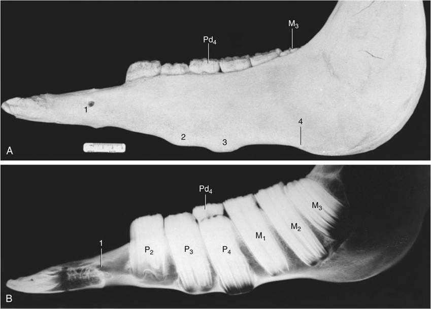
FIG. 18.22 (A) Photograph and (B) radiograph of the left half-mandible of a horse 3 years old (estimated). Note the transitory tubercles on the ventral border and the wedged-in cap (Pd4) that retards the advance of P3 and P4. 1, Mental foramen; 2 and 3, tubercles over the proximal ends of P3 and P4, respectively; 4, notch for facial artery and vein.
The temporalis almost fills the temporal fossa, where it is easily palpated despite the partial covering of thin muscles concerned with the movement of the external ear (Fig. 18.23/1). It arises from the wall of the fossa and from the sagittal crest that forms its median margin, and it envelops the coronoid process of the mandible. On contraction it raises the mandible.
The pterygoideus medialis and lateralis, deep to the mandible, broadly correspond to the masseter in position, orientation, and attachments (Fig. 18.23/2 and 3). The medial muscle, always the larger, extends from the pterygoid process to the mandibular margin.
The lateral muscle runs more horizontally to insert close to the condyle. The masseter and contralateral pterygoid muscles act together to produce the horizontal shifts that supply the principal grinding movement.The digastricus and Occipitomandibularis (strictly a part of the digastricus; Fig. 18.23/4 and 4') are responsible for active opening of the mouth. Despite its much greater bulk, the latter may be regarded as a detachment from the caudal belly of the digastricus. It extends between the paracondylar process of the occipital bone and the caudal border of the mandible. The much more slender digastricus has a similar origin. It presents an intermediate tendon that passes through a split in the insertion of the stylohyoideus. The rostral belly attaches to the ventromedial part of the molar region of the mandible. When the mouth is closed, contraction of the digastricus raises the hyoid apparatus (by virtue of its association with the stylohyoideus) and thus the root of the tongue (Fig. 18.23B).
A thick intra-articular disk is interposed between the expanded and rather flat facets of the mandibular condyle and articular tubercle of the temporal bone (Fig. 18.23A/5). Hinge movements occur at the lower level, which is supported by a tight capsule; the lateral and slight protrusive movements occur at the upper level where the joint cavity is more capacious. The whole joint is supported by a fibrous lateral ligament and an elastic caudal one.
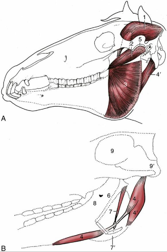
FIG. 18.23 (A) The deep masticatory muscles of the left side have been exposed by removal of the left mandibular ramus (stippled). (B) Medial view of the right digastricus and some related structures. 1, Temporalis; 2, pterygoideus lateralis; 3, lateral surface of pterygoideus medialis; 4, digastricus; 4', occipitomandibularis; 5, left temporomandibular joint; 6, stylohyoid; 7, stylohyoideus; 7', insertion of stylohyoideus on thyrohyoid; 8, medial surface of right mandible and mandibular foramen; 9, cranial cavity; 9', foramen magnum.