THE MUSCLES OF THE FOREARM
The Extensor Group
With one exception—the extensor carpi obliquus—all carpal and digital extensors arise from the craniolateral aspect of the distal end of the humerus and occupy the craniolateral part of the forearm.
Their insertion tendons begin a little above the carpus and are secured in their passage over the joint by condensed deep fascia known as the extensor retinaculum (Figure 23-15, B/77). Each is also individually protected by a synovial sheath, from just above to well below the carpus (Figure 23-16).Except for the ulnaris lateralis, all are extensor to the carpus; the longer muscles also extend the joints of the digit. In addition, their origin provides them with some capacity to flex the elbow, although they are probably little used in this role. All are supplied by the radial nerve. They may each be identified on palpation, and several provide quite conspicuous visible features of the forearm of thin-skinned animals.
The extensor carpi radialis (Figure 23-17/5), the most medial member of the group, runs directly to the front of the subcutaneous border of the radius. Its epimysial covering is joined by the lacertus fibrosus that enables it passively to prevent flexion of the carpal joint when weight is on the limb.
The common digital extensor (Figure 23-17/6) possesses a rather slight radial head in addition to the more substantial origin from the humerus. The radial head is never fully incorporated in the main mass and separates in the lower part of the forearm; its tendon joins that of the lateral digital extensor within the cannon. The main tendon continues down the dorsal aspect to the metacarpus and digit to insert on the extensor process of the distal phalanx. Just before this, it is joined by branches of the interosseus that wind around the sides of the digit from the palmar aspect (Figure 23-17/75).
The slighter lateral digital extensor (Figure 23-17/7) creates a prominent ridge on the lateral aspect of the forearm.
It is joined by the contribution from the common extensor in the upper part of the cannon and then gently inclines toward the dorsal aspect of the limb to insert on the proximal end of the proximal phalanx.The ulnaris lateralis (Figure 23-17/9) runs down the caudal aspect of the forearm. Its short tendon of inser-
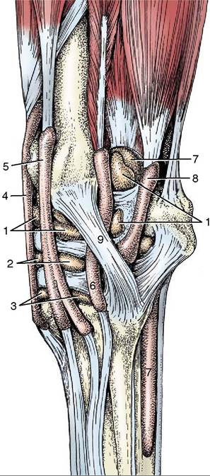
Figure 23-16 Synovial structures of the left carpus; lateral view. 1, Radiocarpal joint capsule; 2, midcarpal joint capsule; 3, carpometacarpal joint capsule; 4, tendon sheath of extensor carpi radialis; 5, tendon sheath of common digital extensor; 6, tendon sheath of lateral digital extensor; 7, tendon sheath of superficial and deep digital flexors (carpal sheath); 8, tendon sheath of ulnaris lateralis; 9, lateral collateral ligament.
tion splits above the accessory carpal bone; a part at once inserts on this bone, while a longer branch descends over the lateral aspect of the bone, tunnels under the collateral ligament, and ends on the head of the lateral splint bone. The longer division requires the protection of a synovial sheath (Figure 23-16/5).
The extensor carpi obliquus is distinguished by arising from the shaft of the radius. It runs in a mediodistal direction to insert on the medial splint bone. Although largely covered by the other muscles, its tendon becomes superficial to that of the extensor carpi radialis (Figure 23-17/5).
The Flexor Group
The muscles of the flexor group also share several attributes. They arise from the caudomedial aspect
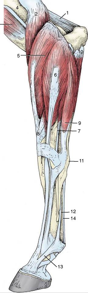
Figure 23-17 Distal muscles of the left forelimb; lateral view. 1, Anconeus; 2, brachialis; 3, biceps; 4, deltoid tuberosity of humerus; 5, extensor carpi radialis; 6, common digital extensor; 7, lateral digital extensor; 8, extensor carpi obliquus; 9, ulnaris lateralis; 10, ulnar head of deep digital flexor; 11, accessory carpal bone; 12, interosseus; 13, extensor branch of interosseus; 14, flexor tendons.
of the humerus, occupy the caudal part of the forearm, obtain their innervation from the median and ulnar nerves, and are flexor to the carpal joint; those that proceed beyond this level are also flexor to the digital joints.
The flexor carpi radialis (Figure 23—18/8) follows the subcutaneous border of the radius and covers the important median vessels and nerve. The tendon of insertion tunnels through the flexor retinaculum where it obtains the necessary protection of a synovial sheath before attaching to the medial splint bone.
The flexor carpi ulnaris (Figure 23-18/9) lies on the medial aspect of the forearm, partly under cover of the flexor carpi radialis. It arises by two heads—from the humerus and the ulna—and inserts on the proximal margin of the accessory carpal bone by means of a short tendon that has no need of synovial protection.
The superficial digital flexor occupies a central position within the flexor group, between the larger mass of the deep flexor and the flexor carpi ulnaris (Figure 23-19/9). A purely tendinous head, usually known as an accessory or check ligament (Figure 23-19/4), arises from the caudal surface of the radius to join the main tendon in the lower part of the forearm; it is a component of the passive stay-apparatus (see further on). The superficial and deep flexor tendons share a common synovial sheath, the carpal sheath, in their passage through the carpal canal.
The tendon is superficial to that of the deep tendon in the metacarpus, but at the fetlock it obtains the deeper position necessary for its insertion on neighboring parts of the proximal and middle phalanges (Figure 23-18/78).
The deep digital flexor is by far the largest of the flexors, although this is not apparent without dissection (Figure 23-19/9'). In addition to the humeral head, there are lesser heads of origin from the upper parts of the radius and ulna. The common tendon passes through the carpal canal and continues down the palmar aspect of the limb to find insertion on the palmar surface of the distal phalanx.
In the metacarpus the tendon is joined by a stout tendinous band that arises from the thick fibrous joint capsule on the palmar aspect of the carpal joint (Figure 23-18/14,14'). This is almost invariably known as an accessory or check ligament; it provides an important element of the passive stay-apparatus that is of far greater significance than the analogous contribution to the superficial tendon.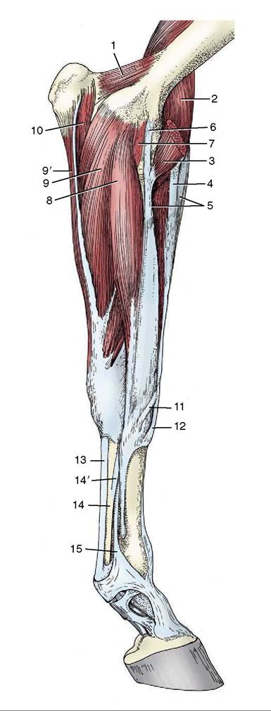
Figure 23-18 Distal muscles of the left forelimb; medial view. 1, Anconeus; 2, brachialis; 3, biceps; 4, lacertus fibrosus; 5, extensor carpi radialis; 6, long part of medial collateral ligament (pronator teres); 7, short part of medial collateral ligament; 8, flexor carpi radialis; 9, 9', humeral and ulnar heads of flexor carpi ulnaris; 10, ulnar head of deep digital flexor; 11, tendon of extensor carpi obliquus; 12, tendon of extensor carpi radialis; 13, tendon of superficial digital flexor; 14, tendon of deep digital flexor; 14', accessory (check) ligament; 15, interosseus.