THE NERVES OF THE HINDLIMB
The lumbosacral plexus and its branches adhere to the common pattern. The obturator nerve (L4-6) crosses the ventral surface of the sacroiliac joint, runs medial to the shaft of the ilium, and passes through the obturator foramen to reach the adductor muscles of the thigh.
It is vulnerable where it lies against bone, and the most common cause of injury is compression during parturition. Conduction is rarely completely interrupted in this injury; cows can still stand and walk on rough ground even when both nerves have been damaged. However, they cannot prevent their feet sliding sideways on smooth floors and, once down, are often unable to rise (Figure 31-12). It must be said that the role of obturator nerve injury in postparturient paralyses (the “downer cow” syndrome) has probably been exaggerated; insufficient attention has been directed toward traumatic or ischemic injury to the adductor muscles ventral to the pelvis as alternative or aggravating causes. These muscles may suffer from direct compression or through constriction of their blood supply in prolonged recumbency.The femoral nerve (L4-6) (Figure 31-11, A) ramifies in the quadriceps after detaching the saphenous branch, which supplies skin over the medial aspect of the limb from midthigh to midmetatarsus. Damage to this nerve is occasionally encountered in newborn calves that were delivered by strong traction on the hindlimbs. An affected limb is unable to bear weight; the diagnosis is confirmed by the loss of sensation in the appropriate area.
Leaving the pelvis, the sciatic nerve (L6-S2) winds around the dorsal and caudal aspects of the hip joint before supplying the caudal muscles of the thigh. Its course between the biceps and semimembranosus, a few centimeters caudal to the femur, exposes it to risk of damage from careless intramuscular injection. Before reaching the gastrocnemius, it divides into tibial and common peroneal nerves, which share responsibility for the innervation of all structures below the stifle, except the medial skin territory of the saphenous nerve.
The sciatic nerve may also be damaged at the birth of an over-large or ill-positioned calf. When the injury is severe, the affected limb hangs loose, and the stifle and hock joints are extended, the digital joints flexed, and the foot knuckled. Cutaneous sensation is lost over most of the extremity.The tibial nerve (L6-S2) passes between the heads of the gastrocnemius and at once detaches branches to the caudal muscles of the leg (Figure 31-11, A), including those that are severed in the treatment of spastic paresis
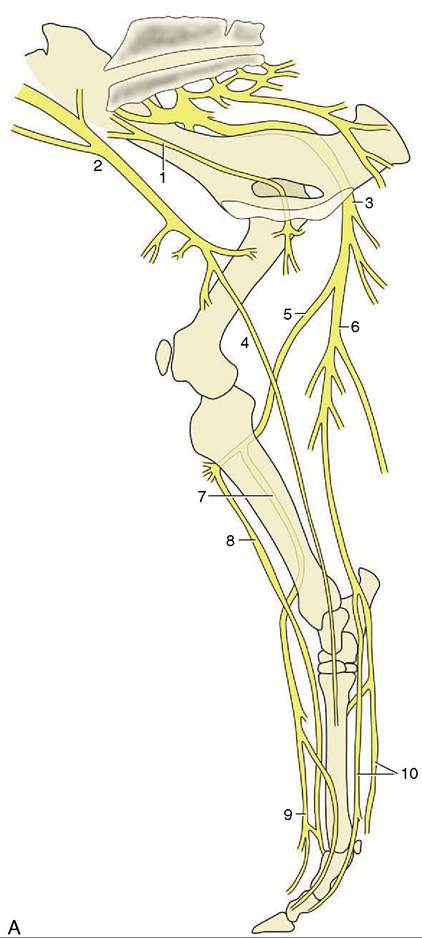
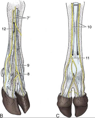
Figure 31-11 Nerves of the right bovine hindlimb. A, Medial view. B, Right hindfoot, dorsolateral view. C, Right hindfoot, plantar view. 1, Obturator n.; 2, femoral n.; 3, sciatic n.; 4, saphenous n.; 5, common peroneal n.; 6, tibial n.; 7, superficial peroneal n.; 7’, lateral and middle branches of superficial peroneal n.; 8, deep peroneal n.; 9, dorsal common digital n. III; 10, medial and lateral plantar nn.; 11, plantar common digital n. III; 12, cranial tributary of lateral saphenous vein.
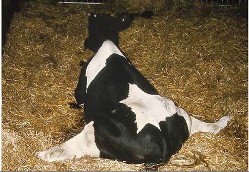
Figure 31-12 Bilateral obturator paralysis.
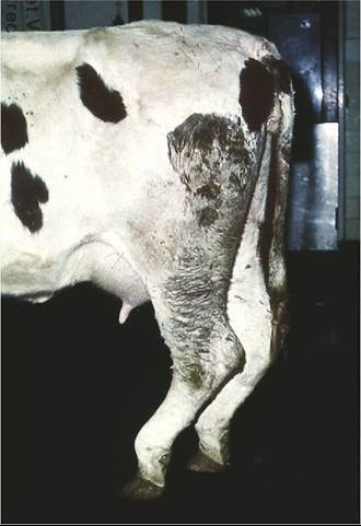
Figure 31-13 Cow with peroneal paralysis.
(see earlier). Severe lesions of this nerve are manifested by overflexion of the hock and overextension of the fetlock, resulting in a vertical pastern. As the digital extensors are not affected, the hoofs are correctly set down as the animal walks and they continue to bear their share of weight at rest. The anomalous attitude of the joints is exaggerated at the walk.
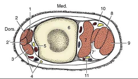
Figure 31-14 Transverse section of the bovine left cannon.
1, Extensor brevis; 2,2', long digital extensor; 3, lateral digital extensor; 4, branches of superficial peroneal nerve and cranial tributary of lateral saphenous vein; 5, deep peroneal nerve and dorsal metatarsal artery (continuation of cranial tibial); 6, metatarsal bone; 7, interosseous; 7’, band from interosseous to superficial digital flexor; 8, deep digital flexor; 9, superficial digital flexor; 10, 11, medial and lateral plantar nerves and vessels.The common peroneal nerve (L6-S2) crosses the gastrocnemius under cover of the biceps to become palpable (and vulnerable) where it passes behind the lateral collateral ligament of the stifle joint. It then sinks between the peroneus longus and the lateral digital extensor before dividing into deep and superficial branches. The larger superficial peroneal nerve crosses deep to the peroneus longus to enter the foot. The deep peroneal nerve supplies the dorsal crural muscles, among which it is embedded, and also enters the foot. Paralysis of the common peroneal is betrayed by overextension of the hock and overflexion of the more distal joints (Figure 31-13).
Unless passively set down correctly, the limb rests on the dorsal surface of the flexed digits. The animal eventually learns to compensate for this defect by flicking the foot forward before placing it on the ground.
The same considerations apply to the digital nerves of the hindfoot as to those of the forefoot. In very brief summary, the dorsal aspect of the foot is the province of the peroneal nerve, and the plantar aspect is the province of the tibial nerve; there is some overlapping to the sides (Figure 31-14).