» The Nerves of the Hindlimb
The formation and ramification of the lumbosacral plexus and the distribution of its peripheral branches follow the common pattern in broad outline; important species differences are confined to the innervation of the foot.
The cranial and caudal gluteal nerves attend to the innervation of the lateral muscles of the croup, including the vertebral heads of the hamstring muscles; the details have been given.
The distributions of the femoral, obturator, and sciatic nerves have greater clinical relevance. The femoral nerve (L4-L6) (Fig. 24.18/1) passes through and also supplies the sublumbar muscles before entering the thigh by way of the vascular lacuna. It then splits into several branches, most of which at once enter the quadriceps. The one branch of more extended course, the saphenous nerve (Fig. 24.18/1'), continues within the femoral triangle before penetrating the medial femoral fascia to obtain a more superficial position. It continues through the thigh, leg, and upper cannon, supplying skin over the medial aspect of the limb from thigh to fetlock. It also supplies the sartorius. Extensive damage to the femoral nerve is uncommon, but when it does occur, the consequences are paralysis of the quadriceps, inability to fix the stifle and therefore inability to support weight on the affected limb, In addition, skin sensibility is lost over a considerable area.
The obturator nerve (L4-L6) (Fig. 24.18/2) leaves the pelvis by way of the obturator foramen and innervates the adductor muscles (pectineus, gracilis, adductor, and obturator externus). Injury, which generally follows foaling or a pelvic fracture, results in partial or complete inability to adduct the limb. The severity of the dysfunction is rather unpredictable depending on the weight of the animal, the nature of the terrain, and the extent of the lesion.
The sciatic nerve (L6-S2) (Fig.
24.18/4) leaves the pelvis by the greater sciatic foramen and, after a short course over the sacrosciatic ligament, turns distally caudal to the hip joint to enter the thigh under cover of the biceps. It divides about the level of the joint into tibial and peroneal nerves that initially run together. They part company a little above the stifle, when the peroneal nerve moves laterally to pass between the biceps and the lateral head of the gastrocnemius. The tibial nerve holds its course and runs between the two heads of the gastrocnemius. Both divisions detach cutaneous branches while still within the thigh. That from the peroneal (lateral cutaneous sural nerve; Fig. 24.18/5') becomes subcutaneous by piercing the biceps and then spreads to supply skin over the lateral aspect of the leg. The corresponding tibial branch (caudal cutaneous sural nerve; Fig. 24.18/6') descends in the fascial plate between the calcanean tendon and deep flexor, following the lateral saphenous vein for part of its course. It supplies branches to the skin over the plantarolateral aspect of the hock and cannon, reaching to the fetlock.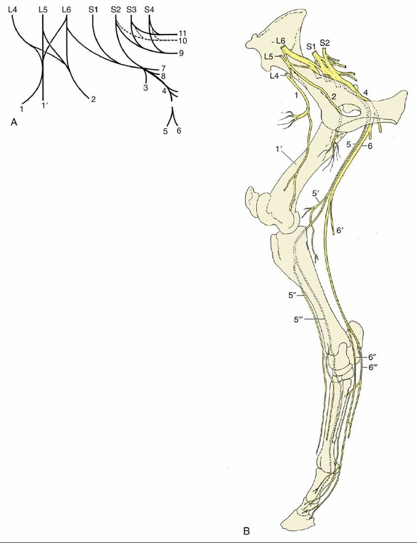
FIG. 24.18 The nerves of the hindlimb. (A) The lumbosacral plexus, schematic. L, Lumbar; S, sacral. (B) The principal nerves, medial view. 1, Femoral nerve (n.); 1', saphenous n.; 2, obturator n.; 3, cranial gluteal n.; 4, sciatic n.; 5, common peroneal n.; 5', lateral cutaneous sural n.; 5" and 5'", superficial and deep peroneal nerves, respectively; 6, tibial n.; 6', caudal cutaneous sural n.; 6" and 6'", medial and lateral plantar nerves, respectively (the lateral nerve gives rise to the plantar metatarsal nerves); 7, caudal gluteal n.; 8, caudal cutaneous femoral n.; 9, pudendal n.; 10, pelvic n.; 11, caudal rectal n.
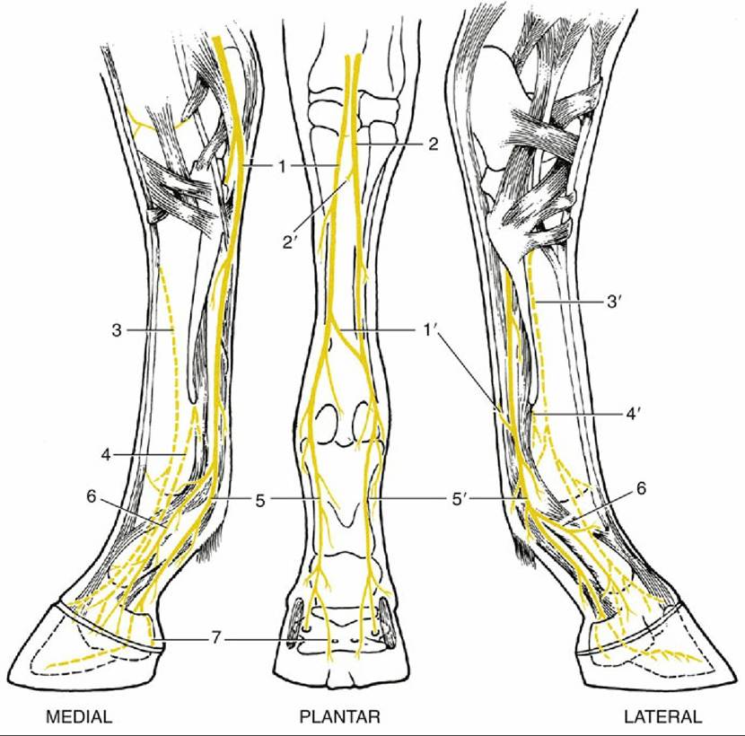
FIG. 24.19 The nerves (nn.) of the right hindfoot.
1 and 2, Medial and lateral plantar nn. (from tibial), respectively; 1', communicating branch; 2', deep branch (for plantar metatarsal nn.), cut; 3 and 3', medial and lateral dorsal metatarsal nn. (from deep peroneal), respectively; 4 and 4', medial and lateral plantar metatarsal nn. (from lateral plantar, 2), respectively; 5 and 5', medial and lateral digital nn., respectively;6, dorsal branch of digital nerve; 7, branch to digital cushion.
The peroneal nerve divides caudal to the lateral collateral ligament of the stifle into deep and superficial branches. The superficial branch (Fig. 24.18/5") continues down the leg, slightly sunken within the groove between the long and lateral extensors, where it can be palpated below the middle of the leg. It supplies the lateral extensor, the skin over the lateral aspect of the leg, and more distal segments of the limb. The deep branch takes a parallel course after sinking deeply between the same two muscles to follow the cranial face of the intervening septum (Figs. 24.18/5'" and 24.8/6'). It supplies branches to the remaining muscles of the dorsolateral group and then continues under cover of the long extensor tendon as a purely sensory nerve that splits into medial and lateral branches over the hock. These, the medial and lateral dorsal metatarsal nerves, edge toward the grooves between the cannon and splint bones (Fig. 24.19/3 and 3'). The lateral nerve follows the palpable dorsal metatarsal artery (Fig. 24.20/8). After detaching twigs to the skin and the fetlock and pastern joints, both finally fade within the hoof.
Complete section of the peroneal nerve results in inability to extend the digit actively. The hoof rests on its dorsal surface unless the ground surface is passively set down. The posture invites comparison with that which occurs in radial paralysis. Afflicted animals may learn to compensate by flicking the foot forward and planting the hoof before the impetus is lost. In addition to the motor disability, skin sensation is lost over the dorsolateral aspect of the lower part of the limb.
Peroneal lesions are most frequent in two circumstances: intrapelvic damage to the sciatic nerve (which is likely also to involve the tibial division) and trauma in the region of the fibula, where the nerve is superficial (see Fig. 31.13, shown on a cow).The tibial nerve dives between the two heads of the gastrocnemius and crosses the stifle on the surface of the popliteus. It detaches branches to these muscles and to other muscles of the caudal group before continuing as a sensory trunk in the space between the calcanean tendon and the deep flexor, where it is easily palpated (Fig. 24.8/12). When level with the calcaneus, it divides into medial and lateral plantar nerves that pass over the sustentaculum tali beside the deep flexor tendon. The lateral nerve diverges laterally, and just distal to the hock, it detaches the common trunk of the medial and lateral plantar metatarsal nerves (Fig. 24.19/2'). These supply the interosseous muscle and associated structures and the plantar pouch of the fetlock joint (Fig. 24.19/4 and 4'). The medial plantar nerve follows the line of the parent trunk. Although the plantar nerves generally resemble the palmar nerves of the forelimb, the communicating branch is relatively slight or even absent; when present, it can usually be palpated as it slopes in a laterodistal direction over the superficial aspect of the flexor tendons (Fig. 24.19/1).
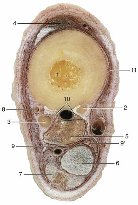
FIG. 24.20 Transverse section of the middle of the left metatarsus. 1-3, Large and small metatarsal bones; 4, long digital extensor; 5, interosseous; 6, deep digital flexor; 7, superficial digital flexor; 8, dorsal metatarsal artery and lateral dorsal metatarsal nerve; 9 and 9', lateral and medial plantar vessels and nerve; 10, plantar metatarsal vessels and nerves; 11, medial dorsal metatarsal nerve.
There is one other difference. The dorsal and plantar metatarsal nerves play a larger role in the sensory innervation of the hoof contents than do the corresponding forelimb trunks—the dorsal branch of the ulnar nerve and the palmar metacarpal nerves — which commonly fail to reach the coronet.
Tibial paralysis is manifested by a slight sagging of the hock when weight is borne on the affected limb. Despite the inability to flex the distal joints, the gait is not seriously disturbed. The sensory deficit is very considerable.
Lesions that affect the sciatic trunk involve the hamstring as well as the leg muscles. Despite this, the consequences are less disastrous than might well be supposed. Retention of activity by the quadriceps enables the animal to fix the stifle and, through the reciprocal apparatus, the hock. It is thus able to support weight on the limb. Cutaneous and deep sensations are lost below the stifle, except in the province of the saphenous nerve.
The tibial nerve may be blocked on the lateral side of the limb approximately 10 cm above the point of the hock.
Both superficial and deep branches of the peroneal nerve can be blocked by injecting, subcutaneously and then deeply from the same point of entry, between the long and lateral extensors a handbreadth or so proximal to the tarsocrural joint (Fig. 24.8/6 and 6'). Apart from this, the local anesthetic techniques for surgical and diagnostic purposes generally resemble those prescribed for the forelimb; the one distinction of relevance is the distal extension of the dorsal metatarsal nerves. It is possible to block the undivided tibial nerve (level with the point of the hock) as an alternative to the plantar nerves (Fig. 24.8/12).
Comprehension Check
Demonstrate the anatomy and mechanics of the reciprocal apparatus.
Compare the core neuromuscular components that ensure the stability of the hindlimb with similar components in the forelimb.