THE NOSE
The nose (nasus) in the broad sense comprises the external nose, the paired nasal cavities, and the paranasal sinuses. A case may be argued for also including the nasopharynx.
An external nose such as forms so conspicuous a feature of the human face is hardly to be recognized in the domestic species, in which it is merged within the general contours of the muzzle (Figure 4-1).
Its extent is more easily determined on palpation as it more or less corresponds with the part of the muzzle skeleton that is cartilaginous and therefore flexible. It is divided internally into two cavities, the nasal vestibules, each of which is entered through a nostril and leads through a region of constriction to the much larger nasal cavity placed beyond. The form and size of the nostrils, their orientation, and the nature of the surrounding integument all show considerable species differences. The integument around the nostrils is naked and sharply demarcated from the unmodified skin in all domestic species other than the horse. According to its extent, the modified region is variously known as the nasal (carnivores, small ruminants), nasolabial (cattle), or rostral (pigs) plate. The nasal plate may be divided by a median groove or philtrum (Figure 4-1/2). The plate is kept moist in cattle, pigs, and dogs; in the first two species the moisture is derived from closely packed underlying glands, whereas in the dog it is an overflow of the secretion of glands of the nasal mucosa, principally the lateral nasal glands.The cartilages that support the external nose are variable in form, relative size, and even number. The rostral end of the nasal septum forms the median partition between the right and left vestibules and includes a small bone (os rostrale) in the pig. The free edge of the septum gives attachment to other cartilages that support the dorsal and lateral margins of the nostril and determine the form of the opening.
One, the alar cartilage, is especially large in the horse and accounts for the curious comma form of the nostril, which is divided in this species into a ventral part, the so-called true nostril leading to the nasal cavity, and a dorsal part, the false nostril leading to a skin-lined diverticulum occupying the nasoincisive notch (see Figure 18-3). The nostril is round in the pig, but in most other species it is prolonged laterally by a slitlike extension. The form of the nostril may be altered, principally by the lateral “wing” (ala) actively being raised by certain facial muscles or passively when the air flow is increased in strenuous breathing or sniffing. These changes can be very pronounced in the horse, leading to compression and almost complete obliteration of the diverticulum.The integument is carried some distance into the vestibule, where it meets the nasal mucosa at a sharply defined line near which several ducts may open. In the horse these include the nasolacrimal (tear) duct, whose opening is very evident on inspection of the vestibular floor of the live animal; the opening is less easily found in other species, either because the tissues are less pliant (cattle) or because it is placed more deeply (dog). The much smaller openings of the long ducts of the serous lateral nasal glands also discharge in this area. This arrangement aids humidification of the incoming air because the acceleration of flow at the constriction favors vaporization of tears and other watery discharge.
The two nasal cavities occupy a large part of the face: they extend caudally to the transverse bony septum at the rostral end of the cranial cavity (Figure 4-2). Their size may be gauged from the conformation of the head, but the first impression is apt to be grossly misleading. Several features greatly reduce the extent of the cavities below expectation. Firstly, certain bones bounding the cavity are thickened by air spaces (paranasal sinuses) that communicate with the cavity but do not form part
of it.
Secondly, the embedded portions of the upper cheek teeth occupy a surprising amount of space, especially in the horse. The potential space is also much reduced by certain very delicate mucosa-covered turbinate bones (conchae) that project into the interior from the dorsal and lateral walls. Finally, the walls are covered by a mucosa locally thickened by vascular plexuses (Figures 4-3, 4-4, and 4-5).The right and left cavities are divided by the nasal septum, which is largely cartilaginous but ossified in its most caudal part (the perpendicular plate of the ethmoid
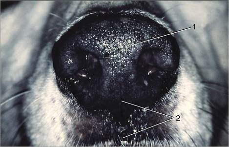
Figure 4-1 The canine muzzle. 1, Nasal plate; 2, philtrum.
bone). The septum meets the upper surface of the hard palate, which separates the nasal and mouth cavities, but the details vary greatly between species (see Figure 4-5). In the horse the septum meets the whole length of the hard palate so that each nasal cavity communicates with the pharynx through a separate opening (choana) (see Figure 18-11). In other species (e.g., ox, dog) the caudal part of the septum fails to meet the palate and a single opening is shared by the two sides (Figures 4-4/7 and 25-9).
The conchae, which intrude on the cavity, have a complicated and variable pattern. Classified by topography (and not by morphology), they comprise a caudal system (of ethmoidal conchae) constituting the lateral mass or labyrinth of the ethmoid bone and a rostral (nasal) system in which large dorsal and ventral (and a much smaller middle) conchae predominate (Figures 4-2 and 25-9). The numerous ethmoidal conchae are separated by narrow clefts (ethmoidal meatuses), and their pattern is most complicated in species that place much reliance on the sense of smell (Figure 4-4/5,6). The dorsal and ventral nasal conchae impose the meatal pattern of the middle and more rostral parts of the cavity. They are formed of fragile laminae coiled on themselves in a manner that varies with the species and the location. Rostrally, the lamina does not recurve to meet itself and thus bounds a recess of the nasal cavity; more caudally the coil meets itself or the lateral nasal
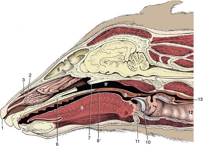
Figure 4-2 Paramedian section of the canine head; the nasal septum has been removed.
1, Right nostril; 2, ventral nasal concha; 3, dorsal nasal concha; 4, ethmoidal conchae; 5, frontal sinus; 6, hard palate; 7, vomer, resected; 8, tongue; 8', oropharynx; 9, soft palate; 9', nasopharynx; 10, epiglottis; 11, basihyoid; 12, trachea; 13, esophagus.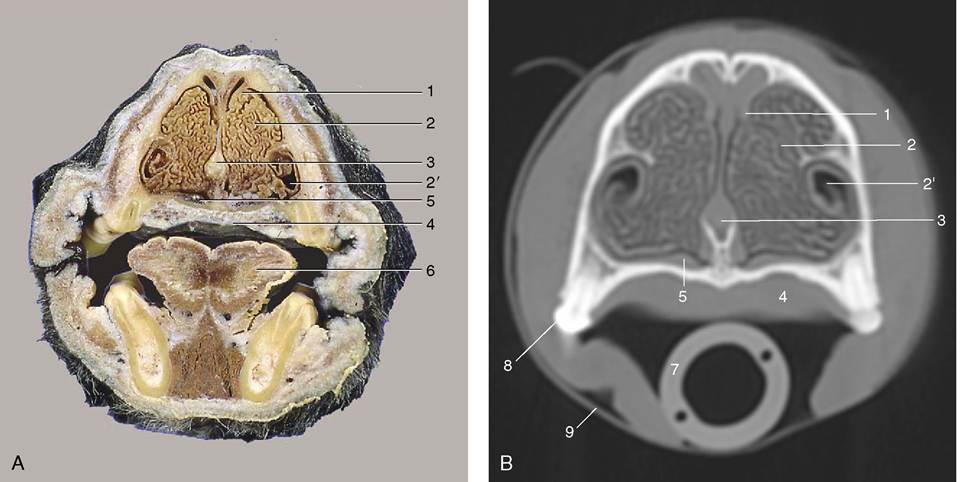
Figure 4-3 A, Transverse section of the canine head at the level of P2. B, CT image taken at the same level but without tongue and structures of the lower jaw. 1, Dorsal concha; 2, ventral concha; 2', recess of ventral concha; 3, nasal septum; 4, hard palate; 5, venous plexus in nasal mucosa; 6, tongue; 7, endotracheal tube; 8, P2; 9, tape to keep endotracheal tube against hard palate during CT procedure.
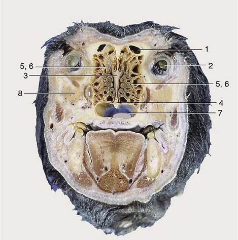
Figure 4-4 Transverse section of the canine head at the level of the eyeball. 1, Frontal sinus; 2, eyeball; 3, ethmoid bone; 4, vomer; 5, 6, ethmoidal conchae; 7, choana; 8, zygomatic gland.
wall to enclose a space that is part of the paranasal sinus system. The conchae reduce the cavity to a series of clefts or meatuses in an arrangement that may be likened to the letter E in transverse section (see Figure 4-5); in other words, the major conchae define dorsal, middle, and ventral meatuses branching from a common meatus against the septum. The dorsal meatus leads directly to the fundus of the nasal cavity and presents air to the olfactory mucosa. The middle meatus usually gives access to the sinus system. The ventral and common meatuses provide the principal airway leading to the pharynx. The relatively wide space at their junction is the route chosen for passage of an instrument such as a stomach tube.
The nasal mucosa blends with the underlying periosteum and varies in thickness. In some parts it is thin, but elsewhere, and especially ventrally, it is much thickened by the inclusion of cavernous blood spaces that make it a semierectile tissue (Figure 4-5Z7, ventral meatus; 8, venous plexus in nasal mucosa.
presented to the lower respiratory passages. The air is warmed by passing over the very vascular mucosa, humidified by the vaporization of the tears and serous nasal secretion, and cleansed by contact with the secretion of numerous scattered mucous glands. These glands spread a carpet of mucus over the nasal mucosa that entraps particles and droplets that come into contact with it. The carpet is moved toward the pharynx by the ciliary action of the lining epithelium and is then swallowed. It is said that in the human species as much as half a liter of mucus is swallowed unconsciously each day.
The paranasal sinuses are diverticula of the nasal cavity that excavate the skull bones (Figure 4-6), largely after birth. The separation of the inner and outer tables of the bones alters the conformation of the head and is especially striking in pigs and cattle (Figures 4-7 and 25-11), in which certain sinuses eventually extend dorsal and even caudal to the cranial cavity. The sinuses retain
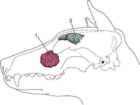
Figure 4-6 Paranasal sinuses in the dog. 1, Maxillary recess; 2, frontal sinus.
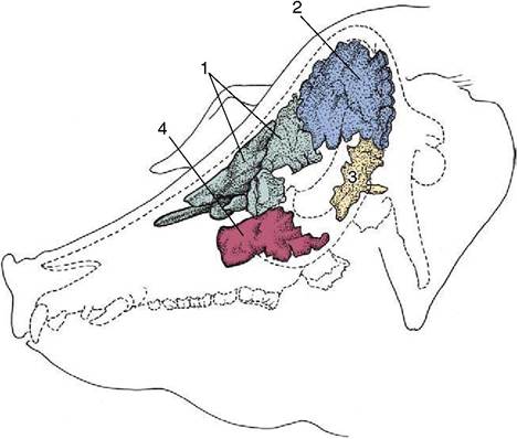
Figure 4-7 Paranasal sinuses in the pig. 1, Rostral frontal sinus; 2, caudal frontal sinus; 3, sphenoidal sinus; 4, maxillary sinus.
their connections with the nasal cavity, but because the openings are generally narrow, a relatively slow exchange of air occurs. The narrowness and locations of the openings make them prone to blockage when the mucosa is thickened by inflammation or congestion. Not all the sinuses are of equal clinical importance; the surface projections of those commonly involved in disease are considered in the topographical chapters.
All species have frontal and maxillary systems, neither communicating with its contralateral counterpart. The frontal system consists of one or more spaces
within the bones at the border between the nasal and cranial cavities.
In most species the various frontal compartments open separately into the ethmoidal meatuses in the nasal fundus, but in the horse the frontal sinus communicates with the nasal cavity indirectly via the caudal maxillary sinus.The maxillary sinus system occupies the caudolateral part of the upper jaw, above the caudal cheek teeth; in some species it sends extensions, variously described as separate sinuses or as diverticula, into the hard palate, the sphenoid bones, the medial aspect of the orbit, and the ventral concha. In the horse the maxillary sinus is divided into caudal and rostral parts, both connected to the middle nasal meatus. In the dog the cavity communicates freely with that of the nose and is known as the maxillary recess.
The function of the sinuses is obscure: they offer some thermal and mechanical protection to the orbit and nasal and cranial cavities, enlarge the skull areas available for muscular attachment without unduly increasing weight, and affect the resonance of the voice.
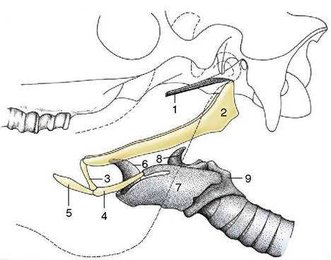
Figure 4-8 Hyoid apparatus suspending the larynx from the base of the skull (horse). The broken line indicates the mandible. 1, Cartilage of auditory tube; 2, stylohyoid; 3, keratohy- oid; 4, thyrohyoid; 5, lingual process of basihyoid; 6, epiglottic cartilage; 7, thyroid cartilage; 8, arytenoid cartilage; 9, cricoid cartilage.