The nose* (nasus) in the broad sense comprises the external nose, the paired nasal cavities, and the paranasal sinuses.
A case may be argued for also including the nasopharynx.
The true extent of the external nose is not readily apparent because it lies within general features of the muzzle; its margins correspond with the cartilaginous and flexible skeleton of the muzzle (Fig.
4.1). The external nose is divided internally into two cavities, the nasal vestibules, each of which is entered through a nostril and leads through a region of constriction to the much larger nasal cavity placed beyond. The form and size of the nostrils, their orientation, and the nature of the surrounding integument all show considerable species differences. The integument around the nostrils, which is naked and sharply demarcated from the unmodified skin in all domestic species other than the horse, is known as the nasal (carnivores, small ruminants), nasolabial (cattle), or rostral (pigs) plate. The nasal plate may be divided by a median groove or philtrum (Fig. 4.1/2). The plate is moist to touch because of the secretions of the underlying glands in cattle and pigs and from secretions of the nasal mucosa, principally the lateral nasal glands, in dogs.Many cartilages support and shape the external nose. The rostral part of nasal septum creates the right and left vestibules and includes a small bone (os rostrale) in the pig. Other cartilages, such as alar cartilage, attach to the free edges of the septum, support the dorsal and lateral margins, and determine the form of the opening of the nostrils. For example, the large alar cartilage creates the unique comma form of the equine nostril, which is divided into a ventral part, the so-called true nostril leading to the nasal cavity, and a dorsal part, the false nostril leading to a skin-lined diverticulum occupying the nasoincisive notch (see Fig. 18.3). The nostril is round in the pig, but in most other species it is prolonged laterally by a slitlike extension.
The form of the nostril may be altered actively by the actions of facial muscles on the lateral "wing" (ala) or passively by the increased airflow in strenuous breathing or sniffing. These changes can be very pronounced in the horse, leading to compression and almost complete obliteration of the diverticulum.The integument meets with the mucosa in the vestibule at a sharply defined line. Near this line the long ducts of the serous lateral glands open along with the prominent opening of the nasolacrimal (tear) duct on the vestibule floor in the horse; the opening is less easily found in other species, either because the tissues are less pliant (cattle) or because it is placed more deeply (dog). This arrangement aids humidification of the incoming air.
The nasal cavities occupy a large part of the face and extend caudally up to the transverse bony septum at the rostral end of the cranial cavity (Fig. 4.2). The use of conformation of the head to estimate the cavities' size is grossly misleading because several features greatly reduce the extent of the cavities to below expectation. First, certain bones bounding the cavity are thickened by air spaces (paranasal sinuses) that communicate with the cavity but do not form part of it. Second, the embedded portions of the upper cheek teeth occupy a surprising amount of space, especially in the horse. Third, the very delicate mucosa-covered turbinate bones (conchae) project into the interior of the cavities from the dorsal and lateral walls. And last, the walls are covered by a mucosa locally thickened by vascular plexuses (Figs. 4.3-4.5).
The right and left cavities are divided by the nasal septum, which is largely cartilaginous but is ossified in its most caudal part (the perpendicular plate of the ethmoid bone). The septum meets the upper surface of the hard palate, which separates the nasal and mouth cavities, but the details vary greatly among species (Fig. 4.5). In the horse the septum meets the whole length of the hard palate so that each nasal cavity communicates with the pharynx through a separate opening (choana) (see Fig.
18.11). In other species (e.g., ox, dog) the caudal part of the septum fails to meet the palate, and a single opening is shared by the two sides (Figs. 4.4/7 and 25.9).The conchae, which are coiled fragile laminae that intrude on the cavity, have a complicated and variable pattern. Classified by topography (and not by morphology), they comprise a caudal system (of ethmoidal conchae) constituting the lateral mass or labyrinth of the ethmoid bone and a rostral (nasal) system in which large dorsal and ventral (and a much smaller middle) conchae predominate (see Figs. 4.2 and 25.9). The numerous ethmoidal conchae are separated by narrow clefts (ethmoidal meatuses) and have a highly complicated pattern in species with a sharp sense of smell (Fig. 4.4/5 and 6). The dorsal and ventral nasal conchae create meatuses of the middle and more rostral parts of the cavity. The form of the conchae varies with the species and the location. Rostrally, the lamina does not recurve to meet itself and thus bounds a recess of the nasal cavity; more caudally the coil meets itself or the lateral nasal wall to enclose a space that is part of the paranasal sinus system. The major conchae define dorsal, middle, and ventral meatuses branching from a common meatus against the septum, and the arrangement looks like the letter E in transverse section (Fig. 4.5). The dorsal meatus leads directly to the fundus of the nasal cavity and presents air to the olfactory mucosa. The middle meatus usually gives access to the sinus system. The ventral and common meatuses provide the principal airway leading to the pharynx. The stomach tube is passed through the wide space at the junction of the meatuses.
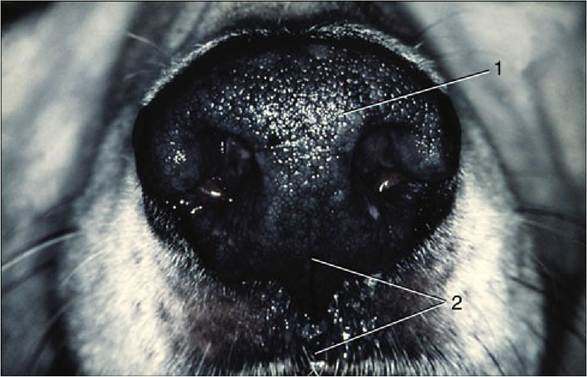
FIG. 4.1 The canine muzzle. 1, Nasal plate; 2, philtrum.
The nasal mucosa blends with the underlying periosteum. In some parts the mucosa is thin, but elsewhere, and especially ventrally, it is much thickened by the inclusion of cavernous blood spaces that make it a semi-erectile tissue (Fig.
4.5/8). The mucosa may become congested and thicker in conditions such as head cold so as to greatly impede the airflow and resulting in stuffiness.Apart from olfaction, the nasal cavity warms the air passing over the highly vascular mucosa, humidified by the vapors of tears and serous nasal secretions, and cleans by interaction with the mucus. The mucous glands create a carpet of mucus on nasal mucosa that entraps particles in the incoming air. The mucous carpet is moved toward the pharynx by the ciliary action of the lining epithelium and is then swallowed. The human species may swallow as much as half a liter of mucus unconsciously each day.
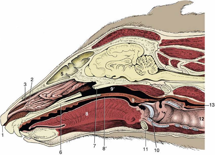
FIG. 4.2 Paramedian section of the canine head; the nasal septum has been removed. 1, Right nostril; 2, ventral nasal concha; 3, dorsal nasal concha; 4, ethmoidal conchae; 5, frontal sinus; 6, hard palate; 7, vomer, resected; 8, tongue; 8', oropharynx; 9, soft palate; 9', nasopharynx; 10, epiglottis; 11, basihyoid;
12, trachea; 13, esophagus.
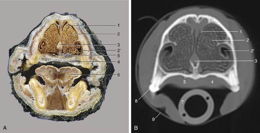
FIG. 4.3 (A) Transverse section of the canine head at the level of P2. (B) Computed tomography (CT) scan taken at the same level but without the tongue and structures of the lower jaw. 1, Dorsal concha; 2, ventral concha; 2', recess of ventral concha; 3, nasal septum; 4, hard palate; 5, venous plexus in nasal mucosa; 6, tongue; 7, endotracheal tube; 8, P2; 9, tape to keep endotracheal tube against hard palate during CT procedure.
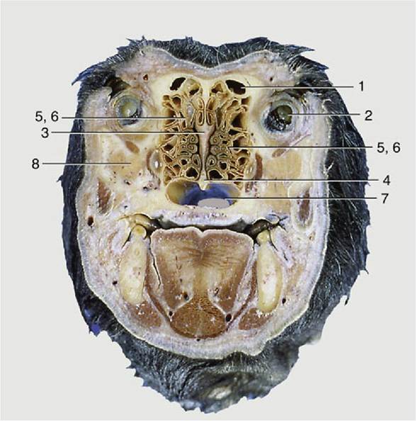
FIG. 4.4 Transverse section of the canine head at the level of the eyeball. 1, Frontal sinus; 2, eyeball; 3, ethmoid bone; 4, vomer; 5 and 6, ethmoidal conchae; 7, choana; 8, zygomatic gland.
The paranasal sinuses are diverticula of the nasal cavity that develop postnatally through the separation of the inner and outer tables of the skull bones (Fig.
4.6). These processes alter the conformation of the head, which is especially striking in pigs and cattle (Figs. 4.7 and 25.11) because certain sinuses do extend dorsal and even caudal to the cranial cavity. The narrow openings of the sinuses slow the exchange of the air with the nasal cavity and are also prone to blockage by congested mucosa in inflammation. Because no all the sinuses are of equal clinical importance, the surface projections of only those commonly involved in disease are considered in the topographical chapters.All species have frontal and maxillary sinus systems, neither communicating with its contralateral counterpart. The frontal system consists of one or more spaces within the bones at the border between the nasal and cranial cavities. In most species the various frontal compartments open separately into the ethmoidal meatuses in the nasal fundus, but in the horse the frontal sinus communicates with the nasal cavity indirectly via the caudal maxillary sinus.
The maxillary sinus system occupies the caudolateral part of the upper jaw, above the caudal cheek teeth. Its extensions into the hard palate, the sphenoid bones, the medial aspect of the orbit, and the ventral concha seen in some species are described as separate sinuses or as diverticula. The horse's maxillary sinus has caudal and rostral parts that are connected to the middle nasal meatus. In the dog the maxillary cavity communicates freely with the cavity of the nose and is known as the maxillary recess.
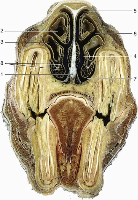
FIG. 4.5 Transverse section of the equine head at the level of P4. 1, Nasal septum; 2, dorsal concha; 3, ventral concha; 4, common meatus; 5, dorsal meatus; 6, middle meatus; 7, ventral meatus; 8, venous plexus in nasal mucosa.
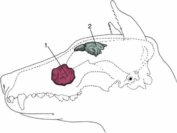
FIG. 4.6 Paranasal sinuses in the dog. 1, Maxillary recess; 2, frontal sinus.
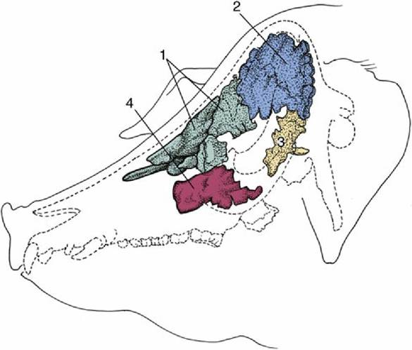
FIG. 4.7 Paranasal sinuses in the pig. 1, Rostral frontal sinus; 2, caudal frontal sinus; 3, sphenoidal sinus; 4, maxillary sinus.
The function of the sinuses is obscure, but they offer some thermal and mechanical protection to the orbit and nasal and cranial cavities, enlarge the skull areas available for muscular attachment without unduly increasing weight, and affect the resonance of the voice.