The Omasum
The omasum lies within the intrathoracic part of the abdomen to the right of the midline, between the rumen and reticulum to the left and the liver and body wall to the right (Figs.
28.7/2 and 28.9/7). It is bilaterally flattened and displays a long convex border that faces dextrocaudally and a much shorter lesser curvature that faces in the opposite direction. The long axis is more or less vertical in the cadaver, but the position and orientation of the living organ alter constantly. Most of the omasum lies under cover of the 8th to 11th ribs, but in cattle the lower pole generally projects onto the abdominal floor below the costal arch (Fig. 28.19/5). Although its position places most of the omasum beyond direct manual reach, the organ may be examined by auscultation and percussion. The lower pole of the omasum has an extensive attachment to the fundic region of the abomasum around the omasoabomasal orifice. Much of its right surface is covered by and partly connected to the lesser omentum (Fig. 28.4C/13).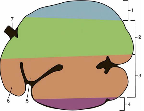
FIG. 28.17 Stratification of ingesta in the ruminoreticulum, left lateral view. 1, Gas bubble; 2, coarse forage (“floating mat”); 3, more finely ground material with higher specific gravity than that in 2; 4, liquid zone; 5, atrium ruminis; 6, reticulum; 7, esophagus.
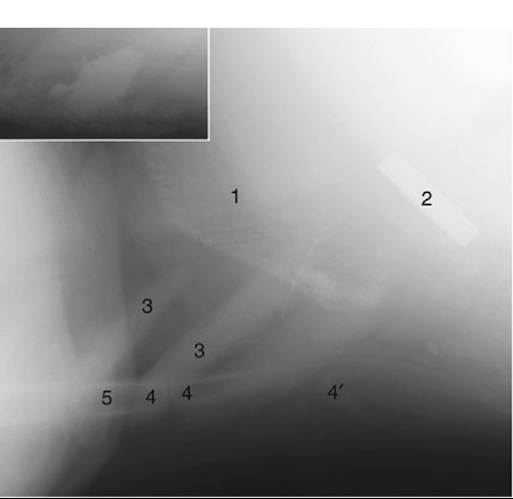
FIG. 28.18 Lateral radiograph of the vicinity of the reticulum of a young cow (cranial is to the left). The inset shows a close-up of a magnet with adhering metal objects. 1, Cranial wall of reticulum with sediment in its “cells”; 2, magnet; 3, costal cartilages; 4, sternebrae; 4', xiphoid cartilage; 5, proximal epiphysis of ulna (olecranon).
The omasum is relatively smaller in sheep and goats, in which it is bean shaped.
It maintains an almost vertical position when the stomach is at rest. It projects on the eighth and ninth ribs but, because of the intervention of the liver, makes no direct contact with the body wall.The interior is occupied by about a hundred crescentic laminae that arise from the sides and greater curvature and project toward the lesser curvature and the omasal canal (see Fig. 28.9). The laminae are of several lengths, and those of different sizes alternate so as to divide the lumen into a series of narrow and fairly uniform recesses (Fig. 28.10/7). The reticulo-omasal orifice is situated at the upper end of the short canal. The large, oval omasoabomasal opening (Fig. 28.9/6) at the other extremity is partly occluded by the prolapse of abomasal folds. The floor of the canal (known as the omasal groove) is smooth except for a few low ridges that run along its length and a scattering of clawlike projections that guard the upper opening.
The keratinized stratified squamous epithelium over the laminae is raised to cover numerous papillae. Most are small and lenticular, but there are a few larger, conical projections that point distally and perhaps promote the onward movement of the ingesta. The mucosa is further characterized by a lamina propria that includes a dense subepithelial capillary network and encloses a thick muscularis mucosae consisting of a thin outer longitudinal layer and a thicker inner circular layer. The inner layer is continuous with the muscle of the omasal wall. The contents of the omasal recesses are finely divided and rather dry, which make the organ firm and easy to recognize on palpation at laparotomy, directly, or from within the rumen after opening that chamber.
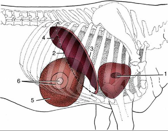
FIG. 28.19 Right lateral projection of certain organs on the bovine thoracic wall. 1, Right atrioventricular valve; 2, position of basal border of lung; 3, cranial extent of diaphragm and liver; 4, field of liver percussion; 5, omasum; 6, field for percussion and auscultation of omasum.
Omasal contractions are biphasic. The first phase squeezes ingesta from the omasal canal into the recesses between the laminae; the second phase is a mass contraction. The principal effect is to squeeze fluid from the material within the recesses, which is a process essential to the continuing movement of ingesta to the abomasum. These contractions occur at a much slower and more deliberate tempo than those of the ruminoreticulum. Although the rough surfaces and muscular cores of the laminae suggest that these folds triturate the food by rubbing against each other, there is no evidence of such activity. Absorption is continued in the omasum.