THE OVARY AND UTERINE TUBE
The ovaries, about 5 cm long, are distinguished by the many follicles and corpora lutea that project from the entire surface (Figure 35-2). They are usually found hidden among the intestines, slightly ventrolateral to the pelvic inlet.
The relatively long mesovaria commonly allow both ovaries to lie against the one flank, and consequently, both may be removed through a single incision.The uterine tube (Figure 35-3Z4) is about 20 cm long and is carried in the wall of the cone-shaped ovarian bursa; it meets the horn of the uterus at a tapering junction. Obstruction of the tube (the origin of hydrosalpinx) can cause infertility in sows.
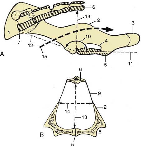
Figure 35-1 A, Median section of the sow's pelvis. B, Transverse section of the pelvis near the level of the vertical diameter. 1, Coxal tuber; 2, ischial spine; 3, ischial tuber; 4, obturator foramen; 5, pelvic symphysis; 6, S4; 7, promontory; 8, acetabulum; 9, sacrosciatic ligament; 10, angle between pelvic floor and conjugata; 11, plane of pelvic floor; 12, conjugata; 13, vertical diameter; 14, transverse diameter; 15, pelvic axis.
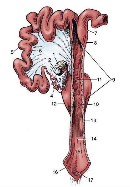
Figure 35-3 The reproductive tract of the sow opened dor- sally in part; the right uterine horn and ovary are not shown. 1, Left ovary; 2, ovarian bursa; 3, mesosalpinx; 4, uterine tube; 5, uterine horn; 6, broad ligament; 7, parallel segments of uterine horns; 8, body of uterus; 9, cervix; 10, external uterine orifice; 11, mucosal prominences; 12, bladder; 13, vagina; 14, external urethral orifice; 15, vestibule; 16, vulva; 17, glans of clitoris.
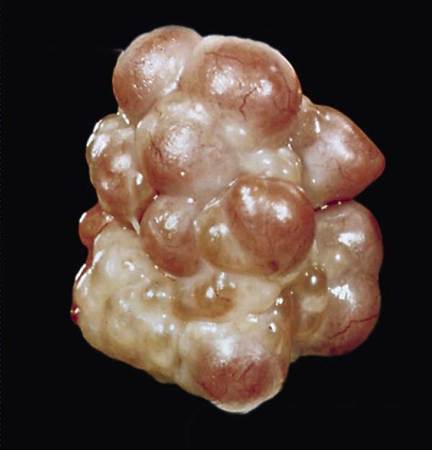
Figure 35-2 Ovary (sow) exhibiting mature follicles.