The pancreas is a much smaller gland closely related to the duodenum in the dorsal part of the abdominal cavity.
It is yellowish and bears some resemblance to the salivary glands, although it is softer and more loosely knit. It combines exocrine and endocrine functions.
The exocrine component, by far the larger, produces a digestive juice that is discharged into the proximal part of the duodenum through one or two ducts.
The juice contains enzymes that break down protein, carbohydrates, and fats. The endocrine component comprises the pancreatic islets, which are cell clumps that are scattered between the exocrine acini and are the source of insulin, glucagon, and gastrin. The islets are therefore of prime importance in carbohydrate metabolism (p. 208).The pancreas is conventionally regarded as consisting of a body and two lobes, a description that suits the canine pancreas but is less apt for those of some other species (Fig. 3.56). When hardened in situ, the canine pancreas is acutely flexed: the apex of the V nestles close to the cranial flexure of the duodenum. The slender right lobe runs within the mesoduodenum. The thicker but shorter left lobe extends over the caudal surface of the stomach toward the spleen, within the greater omentum (see Fig. 3.33/7).
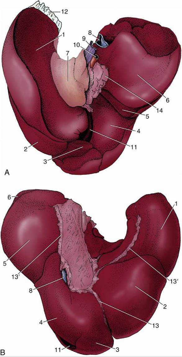
FIG. 3.53 (A) Visceral and (B) diaphragmatic surfaces of the canine liver. 1, Left lateral lobe; 2, left medial lobe; 3, quadrate lobe; 4, right medial lobe; 5, right lateral lobe; 6, caudate process (of caudate lobe); 7, papillary process (of caudate lobe); 8, caudal vena cava; 9, portal vein; 10, hepatic artery; 11, gallbladder; 12, left triangular ligament; 13, falciform ligament; 13', coronary ligaments; 14, lesser omentum.
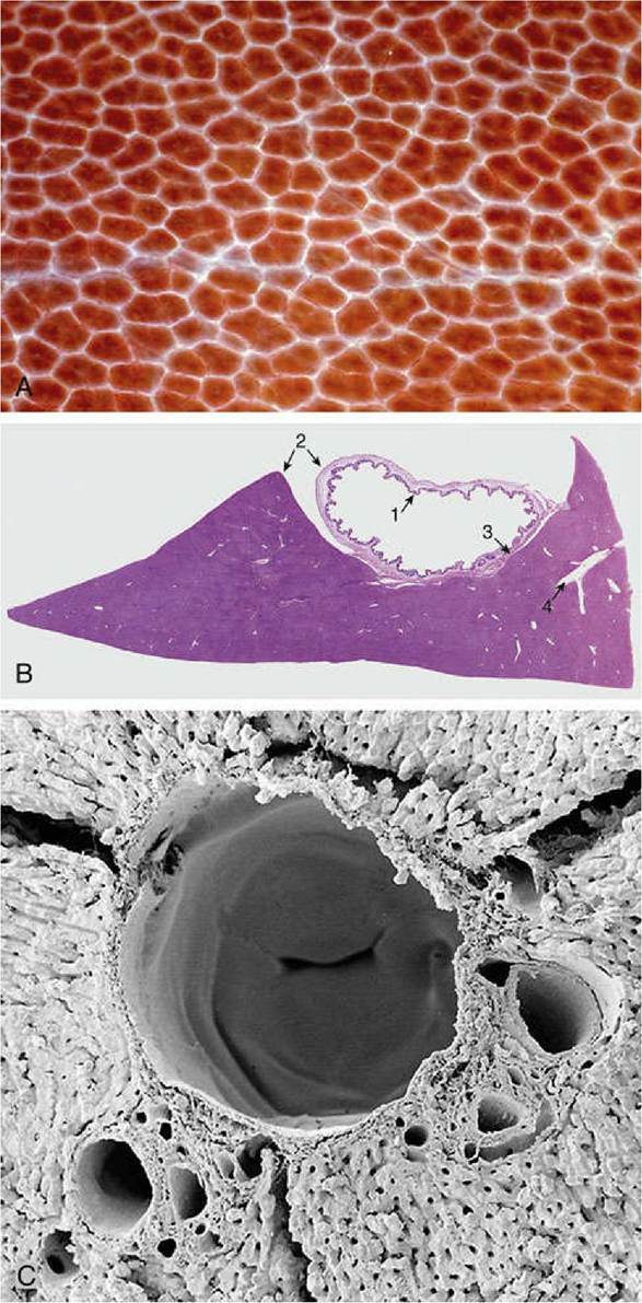
FIG. 3.54 (A) Surface of liver (enlarged) with clearly defined hepatic lobules (pig).
(B) Liver andgallbladder (monkey) (hematoxylin and eosin stain). 1, Tunica mucosa of gallbladder; 2, visceral peritoneum covering the liver and gall bladder surface; 3, tunica adventitia of gallbladder; tunica muscularis is very thin; 4, hepatic portal vessels. (C) Scanning electron microscope image of a corrosion cast of hepatic vessels (rat); note the valve within the central vein.
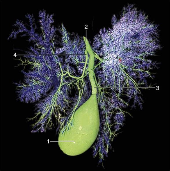
FIG. 3.55 The bile drainage system of the dog. 1, Gallbladder; 2, bile duct; 3, cystic duct; 4, hepatic ducts.
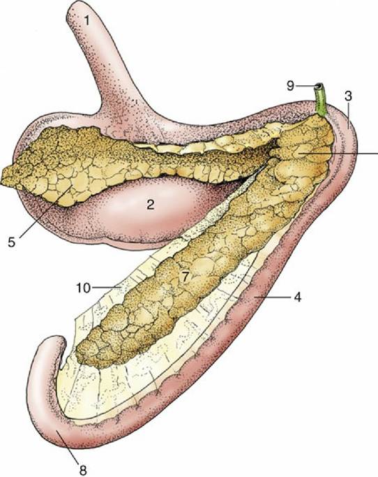
FIG. 3.56 The pancreas of the dog (caudal view). 1, Esophagus; 2, stomach; 3, cranial flexure of duodenum; 4, descending duodenum; 5, left lobe of pancreas; 6, body; 7, right lobe; 8, caudal flexure of duodenum; 9, bile duct; 10, mesoduodenum.
The pancreas arises from two primordia that bud from the proximal part of the duodenum. The buds later merge, but in many species evidence of the dual origin of the pancreas is provided by its duct system. A greater pancreatic duct commonly drains the part of the pancreas that arises from the ventral primordium and opens into the duodenum together with, or just beside, the bile duct. A lesser (accessory) duct emerges from the part of the pancreas formed by the dorsal primordium and opens on the opposite aspect of the gut. This is the arrangement usually found in the dog, although the terminal part of one duct sometimes regresses. Because the duct systems of the two lobes communicate within the gland, the absence of one or the other outlet is of no significance. In some species only one duct commonly survives.
The generous blood supply is from the cranial and caudal pancreaticoduodenal arteries, of which the former branches from the celiac artery and the latter from the cranial mesenteric artery. The veins drain to the portal vein. The gland is supplied by both sympathetic and parasympathetic nerves.