THE PELVIC CAVITY
The pelvic cavity of the cow becomes progressively narrower between the entrance and the exit. It also loses depth but in less regular fashion because a pronounced dip of the middle part of the floor results in a local increase in height before the caudal part slopes steeply upward to the shallow exit (Figure 29-1).
The entrance faces ventrocranially at an angle that carries the pecten of the pubis below the second inter- sacral joint (Figure 29-1/15). Behind the iliac shaft, the width is reduced by inflection of the high ischial spine, and it becomes further reduced by the encroachment of the massive ischial tuber on the exit (Figure 29-2). The conspicuously cramped exit is roughly triangular; the third caudal vertebra and the tubercular ischial tubers are its corners. The lateral border is completed by the sacrotuberous ligament (the edge of the sacrosciatic ligament), while the caudal margin of the floor is cut away at the ischial arch. The strong development of the ischial crest and tuber combine to reduce the contribution to the lateral wall that is made by the sacrosciatic ligament (Figure 29-2/4).
There are certain variations associated with age and gender. The entrance is almost uniformly wide in mature cows but considerably narrowed in its ventral part in heifers. In these younger animals the cranial part of the floor raises a ridge over the symphysis; in older cows, especially those that have carried several calves, the same region is level or sunken. The male girdle, despite being significantly more robust, encloses a cavity that is clearly less capacious; it is even more confined at the entrance, and beyond this the cranial part of the floor tends to be domed.
In sheep and goats the long, slender iliac shafts approach the vertebral column at an acute angle that, in combination with the shortness of the sacrum, places the pecten below the second joint of the tail (see Figure 26-2).
The sacroiliac joints (Figure 29-3) are complemented by strong ligaments binding the two bones together; virtually no movement is normally allowed. About the time of parturition there is some hormone-induced slackening of the collagenous structures of the pelvis, and a modest but potentially significant mobility may then become possible (p. 214).
Ankylosis of these joints, accompanied by lumbar spondylosis, is common in aging bulls and, when severe, may disable the animal for service.
The perineal region is extensive because those parts of the hamstring musculature that in the horse provide it with very prominent lateral boundaries are lacking in cattle.
By convention, the region is considered to extend ventrally to include the nearest part of the udder (or scrotum). The increase in breadth exposes the sacrotu- berous ligaments, the ischial tubers, and the ischiorectal fossae as visible and palpable surface landmarks. The anus and vulva, the most obvious features of the dorsal and ventral perineal regions, respectively, are considered later (see Figure 29-10).
The blood supply to pelvic structures is delivered by the small median sacral artery and the much larger, paired internal iliacs (Figure 29-4). The first or, more accurately, its continuation as the median caudal artery has already been encountered (p. 668). The internal iliac artery serves both parietal and visceral structures, contrary to the usual arrangement. It enters the pelvic cavity close to the sacroiliac joint and continues down the ilium to reach the vicinity of the lesser sciatic foramen (Figure 29-1/10) before dividing into internal pudendal and caudal gluteal arteries. The latter, like other parietal branches, is of no present concern. The internal iliac’s first visceral branch, detached close to the origin of the parent trunk, is the umbilical artery. This term, though appropriate to its role in the fetus, is misleading because the vessel is now almost exclusively concerned with supplying blood to the uterus through a large uterine artery; the continuation of the umbilical, reduced to a fibrous cord with a vestigial lumen, is better known as the round ligament of the bladder.
The male homologue of the uterine artery is the deferential. (The distribution of the arteries to the viscera is considered with the organs they supply.) The second visceral branch, the vaginal artery, is detached close to the termination of the internal iliac trunk and supplies the bulk of the pelvic viscera. The male homologue is the prostatic artery. The internal pudendal artery supplies both parietal structures, including the muscles of the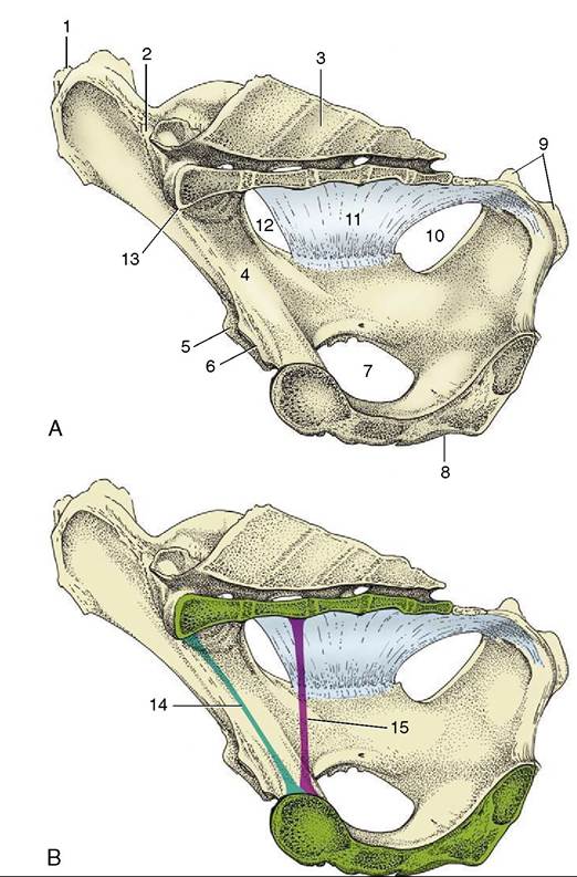
Figure 29-1 A-B, Median section of the bony pelvis of a cow. Certain obstetrical terms are illustrated in B. 1, Coxal tuber; 2, sacroiliac joint; 3, sacrum; 4, shaft of ilium; 5, cranial border of acetabulum; 6, pecten pubis; 7, obturator foramen; 8, symphysis; 9, ischial tuber; 10, lesser sciatic foramen; 11, sacrosciatic ligament; 12, greater sciatic foramen; 13, promontory; 14, conjugate—the line connecting the promontory with the pecten; 15, vertical diameter—the vertical line between the pectin and the pelvic roof.
pelvic diaphragm, and viscera, including the female tract from the caudal vagina to the vestibule. The depleted trunk leaves the pelvis, through an opening in the fascia directly above the symphysis, to supply branches to the clitoris and labia and other branches to the perineum, some of which reach the caudal part of the udder (or scrotum and prepuce).
The nerves within the pelvis fall into two groups (Figure 29-5). The first comprises the obturator and sciatic nerves that, despite their vulnerability to injury at parturition, will be described with the hindlimb. The
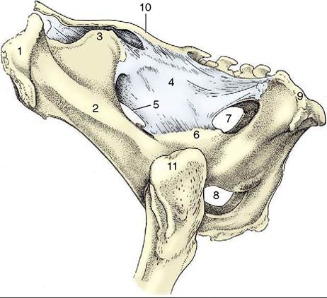
Figure 29-2 Lateral view of the bony pelvis of a cow. 1, Coxal tuber; 2, shaft of ilium; 3, sacral tuber; 4, sacrosciatic ligament; 5, greater sciatic foramen; 6, ischial spine; 7, lesser sciatic foramen; 8, right and left obturator foramina; 9, ischial tuber; 10, sacrum; 11, greater trochanter.
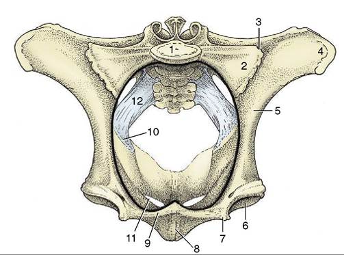
Figure 29-3 Cranial view of the bony pelvis of a cow. The terminal line (black) is indicated. 1, Body of first sacral vertebra; 2, wing of sacrum; 3, sacroiliac joint; 4, coxal tuber; 5, shaft of ilium; 6, acetabulum; 7, iliopubic eminence; 8, symphysis; 9, pecten pubis; 10, ischial spine; 11, obturator foramen; 12, sacrosciatic ligament.
second group comprises the pudendal, caudal rectal, and pelvic nerves, of which all are purely sacral in origin and concerned with the supply of the pelvic viscera and the perineum. The significant divisions of the pudendal nerve are the deep perineal and distal cutaneous branches and the continuation of the main trunk. The deep perineal supplies both visceral and somatic struc-
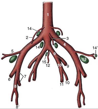
Figure 29-4 Branching pattern of the caudal part of the bovine abdominal aorta. 1, Aorta; 2, ovarian artery; 3, caudal mesenteric artery; 4, external iliac artery; 5, deep circumflex iliac artery; 6, femoral artery; 7, deep femoral artery; 8, puden- doepigastric trunk; 8', external pudendal artery; 9, internal iliac artery; 10, umbilical artery; 11, uterine artery; 12, median sacral artery; 13, deep inguinal (iliofemoral) lymph node; 14, 14’, medial and lateral iliac lymph nodes; 15, sacral lymph nodes.
ally decreasing further until it eventually disappears (Figures 29-7 and 29-8), which brings the rectum into broad contact with the pelvic roof. In this process more and more of the rectal circumference becomes denuded of serosa until the last part is completely embedded in fat, which provides the cushion that allows the gut to adjust to changing circumstances. The close connection with the pelvic roof and walls is a handicap to rectal explorations, and for many purposes the hand must be carried forward into the more mobile colon (Figure 29-9) (p. 720).
The anal canal is embraced by the pelvic diaphragm; the postdiaphragmatic part forms a low eminence presenting a short transverse slit through which the skin continues to provide the last stretch of the canal with a cutaneous epithelial covering. The anus is guarded by the usual two sphincters, and the striated external one exchanges fascicules with other muscles of the perineum (Figure 29-10).
Most of the rectum is supplied from the cranial rectal artery, a branch of the caudal mesenteric, but the terminal section and the anal region are supplied by twigs from the caudal rectal, an indirect branch of the vaginal artery. The venous drainage is divided between the portal and systemic systems.