THE PELVIC REPRODUCTIVE ORGANS
The deferent ducts take their usual courses to penetrate the body of the prostate before opening into the urethra on the summit of a low papilla (Figure 35-8/5). They do not expand to form ampullae and in the last part of their courses are covered by the very large vesicular glands that open beside them (Figure 35-8/7).
Only small parts of these glands are contained within the pelvic cavity; the bulk protrudes into the abdomen, beyond the neck of the bladder (Figure 35-6/7), and is enclosed within the genital folds. In addition to a modest irregular body, the prostate (Figure 35-8/8) possesses a large disseminate part spread within the wall of the pelvic urethra.The bulbourethral glands are remarkable for their shape and size. They lie dorsolateral to the pelvic urethra and are sufficiently long to touch the vesicular glands
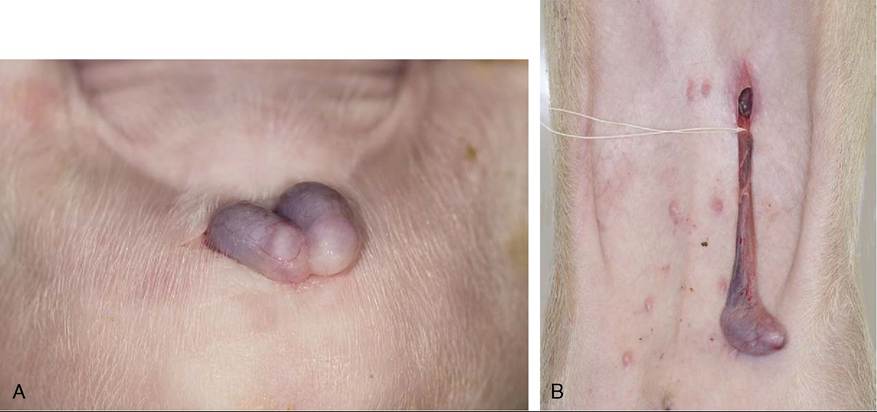
Figure 35-7 A, Open castration method of a newborn piglet. (Note: The parietal layer of vaginal tunic is still intact.) B, The closed castration method in a 5-week-old piglet (also performed in case of an inguinal hernia).
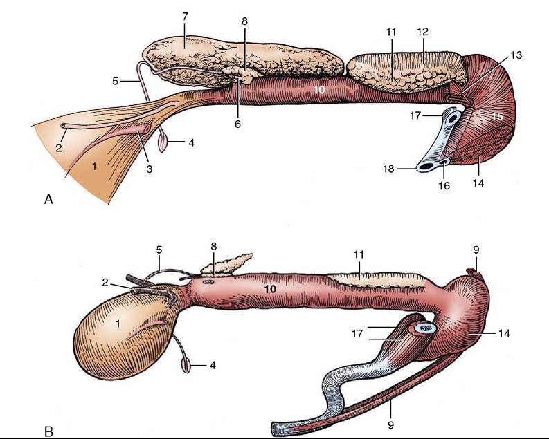
Figure 35-8 Pelvic urethra and associated organs of an 8-month-old boar (A) and a 6-month-old castrate (B), left lateral views. The left vesicular gland has been removed to expose the prostate. 1, Bladder; 2, left ureter; 3, left umbilical artery; 4, right vaginal ring; 5, right deferent duct; 6, left deferent duct, cut at prostate; 7, right vesicular gland; 8, body of prostate; 9, retractor penis; 10, pelvic urethra, surrounded by urethralis; 11, left bulbourethral gland; 12, bulboglandularis covering dorsal half of bulbourethral gland; 13, excretory duct of left bulbourethral gland; 14, bulbospongiosus; 15, bulb of penis; 16, urethra and corpus spongiosum; 17, right and left crura, cut; 18, corpus cavernosum.
(Figure 35-8, AJ11 and Figure 35-6/8). Each drains through a dilated sometimes duplicated duct that opens onto the thickening that separates a dorsal diverticulum from the lumen of the urethra where this bends around the ischial arch. The glands are covered by the bulbo- glandularis muscles, whose contraction secures their evacuation (Figure 35-8, A/12). The caudal ends of the glands may be palpated per rectum. The ability to touch the urethra between them is diagnostic of the castrate (Figure 35-8, B); inability to do this in the absence of palpable testes suggests cryptorchidism.