The Pelvic Reproductive Organs
The short (≈12 cm) pelvic urethra lies directly over the pelvic symphysis. Although generally remarkably wide (≈6 cm), its lumen is narrowed in two places: one level with the body of the prostate, and the other where the urethra crosses the ischial arch (Fig.
22.20). The deferent ducts (Fig. 22.20/2) penetrate the urethral wall close to the origin of the urethra from the bladder. Each combines with the duct of the neighboring vesicular gland to form a common passage, the ejaculatory duct. This is only a few millimeters long and opens into the urethra to the side of the dorsal thickening, the seminal colliculus.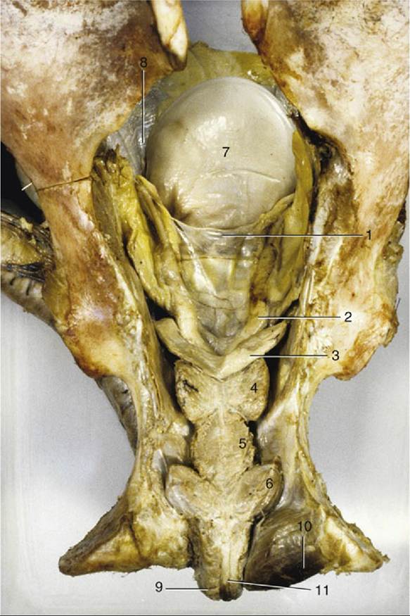
FIG. 22.20 Dorsal view of the pelvic urethra and accessory reproductive glands (in situ). 1, Genital fold; 2, ampulla of deferent duct; 3, vesicular gland; 4, prostate; 5, urethralis; 6, bulbourethral gland; 7, bladder;
8, lateral ligament of bladder; 9, bulbospongiosus; 10, ischiocavernosus; 11, retractor penis.
The vesicular glands (Fig. 22.20/3) of the horse merit the alternative name seminal vesicles because they take the form of smooth-surfaced, pear-shaped bladders, approximately 12 cm long, with large central lumina. Each is contained within the genital fold.
The prostate (Fig. 22.20/4) is largely retroperitoneal and entirely compact. It consists of two lateral lobes joined by a narrow isthmus that crosses the dorsal aspect of the urethra close to the bladder neck. Each lateral lobe is pressed against the border of the urethra and extends cranially along the caudolateral edge of the adjacent vesicular gland. Because the prostate is firm and lobulated, the two glands are easily distinguished on rectal examination. Numerous ductules drain from the prostate to discharge into the urethra through tiny slits beside the colliculus (see Fig.
5.50/7).The paired bulbourethral glands lie dorsolateral to the urethra at the pelvic outlet. They are thinly covered by striated muscle (bulboglandularis), about 4 cm long, and so oriented that their pointed caudal ends converge (Fig. 22.20/6). These glands discharge through numerous small pores that open into the urethra where it leaves the pelvis.
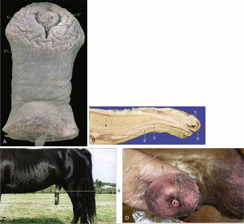
FIG. 22.21 (A) Extremity of penis exposed. (B) Enlarged glans penis. (C) Within prepuce in median section. (D) Enlarged glans penis. 1, Glans; 1', corona glandis; 1", collum glandis; 2, urethra; 2', corpus spongiosum; 3, urethral process within fossa glandis; 3', urethral sinus; 4, corpus cavernosum; 5, preputial fold; 5', preputial ring; 6, prepuce, forming preputial orifice with the body wall.
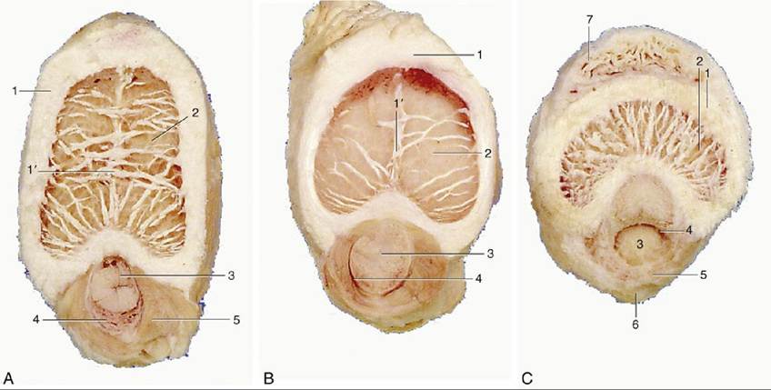
FIG. 22.22 Transections of the penis, (A) directly distal to the root, (B) midshaft, and (C) in its free part.
1, Tunica albuginea; 1', incomplete septum penis; 2, corpus cavernosum; 3, urethra; 4, corpus spongiosum; 5, bulbospongiosus; 6, retractor penis; 7, dorsal process of glans.
All accessory reproductive glands are of course much reduced in geldings.