THE PENIS AND PREPUCE
The penis is suspended below the trunk and is partly contained between the thighs, where it is anchored to the floor of the pelvis by a suspensory ligament in the large species. In the quiescent state, the free extremity is concealed within an invagination of the abdominal skin, the prepuce, which opens at a variable site behind the umbilicus.
The organ is mainly constructed of three columns of erectile tissue (Figure 5-53). These are independent caudally where they constitute the root of the penis, but their major parts are combined in the body of the penis.The paired dorsal columns are known as the crura of the penis (Figure 5-53/7) at their widely separated origins from the ischial arch. They converge, bend forward, and run below the pelvic floor before joining. Each consists of a core of cavernous tissue enclosed
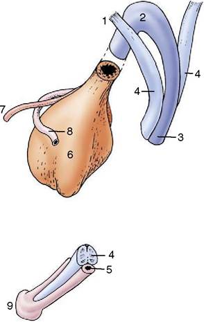
Figure 5-53 Schematic drawing of the components that constitute the equine penis at its root and at its apex. 1, Crus penis; 2, bulb; 3, corpus spongiosum; 4, corpus cavernosum; 5, urethra; 6, bladder; 7, ureter; 8, deferent duct; 9, glans.
within a thick connective tissue casing (tunica albuginea), and the complex is known as a corpus cavernosum (Figure 5-53/4). A septum exists between the two corpora cavernosa in the proximal part of the body, but in most species this will be found to weaken and ultimately disappear when traced distally toward the apex of the penis. In carnivores the septum is complete. The combined structure is grooved ventrally to accommodate the third component, the urethra within its enveloping vascular sleeve, the corpus spongiosum (Figure 5-53/3). The blood spaces within the crura and corpus cavernosum communicate freely.
The corpus cavernosum does not extend to the apex of the penis, which is formed by an expansion of the corpus spongiosum.
The corpus spongiosum commences at the pelvic outlet with the sudden enlargement of the meager spongy tissue of the pelvic urethra. The expansion constitutes the bulb of the penis (Figure 5-53/2), a bilobed structure that tapers to continue as a more uniform sleeve. The corpus spongiosum is more delicate than the corpus cavernosum, having larger blood spaces separated by thinner septa. Its cranial expansion over the distal end of the corpus cavernosum, usually known as the glans (Figure 5-53/9), forms the apex of the whole organ. Since the corpus spongiosum surrounds the urethra, the urethral orifice is brought to the very extremity of the penis; indeed, in small ruminants a free urethral process prolongs the urethra well beyond this.Other pronounced species differences in penis structure exist. In the dog and cat the distal part of the corpus cavernosum is transformed into bone, the os penis. The glans has very different forms. It is minimally developed in the pig, insubstantial in the ruminants, but large and mushroom-shaped in the horse. It is most specialized in the dog, in which it presents bulbar proximal and long cylindrical distal parts. The penis of the cat is unique (among domestic species) in pointing cau- doventrally from the ischial arch; this retention of the embryonic posture affects the manner of copulation.
The construction of the corpus cavernosum also exhibits major differences. In some species it contains small blood spaces enclosed within and divided by substantial amounts of tough fibroelastic tissue. Relatively little additional blood need be retained to make this fibroelastic type of penis become erect (Figure 5-54, A); this construction is found in the penis of the boar and ruminant species in which the quiescent organ exhibits a sigmoid flexure of that part of its body carried between the thighs. In the other type, the blood spaces are relatively larger, and the enclosure and intervening septa more delicate and more muscular (Figure 5-54, B); a relatively much greater quantity of blood is required to achieve erection, which involves significant increases in both length and girth.
This musculocavernous type of penis is found in the stallion and, in atypical form, in the dog.The prepuce or sheath is a tubular fold consisting of an external layer (lamina externa), continuous with the general integument, and an internal layer (lamina interna) that faces the free end of the penis; the internal layer continues as the covering of the free part of the penis after reflection in the depth of the preputial cavity. Both the internal layer and the penile covering are hairless but often well provided with smegma-secreting glands and lymphoid tissue. In the newborn male the penis and sheath are fused, and separation is gradually achieved during the period before puberty (p. 719). The attachments of the adult prepuce are sufficiently loose to allow the internal lamina to be reflected onto the erect penis when this is protruded through the preputial orifice.
Certain muscles are associated with the penis. The bulbospongiosus is the thick extrapelvic continuation of the urethralis. It begins abruptly and extends distally to end on the surface of the corpus spongiosum at a variable distance beyond the point at which this is incorporated within the penis.
The powerful paired ischiocavernosi arise from the ischial arch, almost enclose the crura, and follow them to their fusion.
The retractor penis is also paired. It arises from the caudal vertebrae and descends through the perineum, bending laterally to pass around the anal canal, to reach
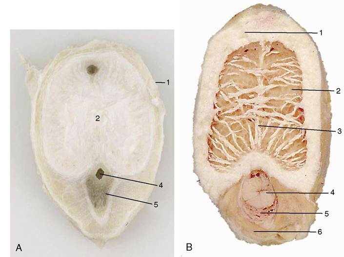
Figure 5-54 Transverse sections of the fibroelastic penis of a bull (A) and the musculocavernous penis of a stallion (B). 1, Tunica albuginea; 2, corpus cavernosum; 3, septum; 4, urethra; 5, corpus spongiosum; 6, bulbospongiosus.
the penis. Unlike the other muscles associated with the penis, the retractor is mainly composed of smooth muscle fibers.
Narrow slips of striated muscle (cranial and caudal preputial) may pass onto the prepuce and attach near its opening.
The caudal muscles are less frequently encountered and retract the prepuce, thus uncovering the extremity of the penis. The cranial muscles protract the prepuce. Both caudal and cranial muscles must be regarded as detachments of the cutaneous trunci; they are best developed in the bull but lacking in the stallion.The penis obtains its exclusive (in the horse, principal) blood supply from the artery of the penis, a terminal branch of the internal pudendal. The artery of the penis has a very short course, and at the ischial arch it quickly divides to form an artery of the bulb, which enters the bulb of the penis and supplies the corpus spongiosum; a deep artery, which pierces the tunica albuginea to supply the corpus cavernosum; and the dorsal artery, which passes apically on the dorsal border of the organ to supply the free end. The dorsal artery may be reinforced by anastomosis with the obturator artery (horse) and generally by anastomosis with the external pudendal artery for the supply of the prepuce. The veins are broadly satellite. Interspecific details are considered in the later chapters when they are significant.
The nerves to the penis accompany the vessels. The motor fibers are predominantly parasympathetic and from the pelvic nerves.