THE PHARYNX
The pharynx is divided in the customary fashion.
The nasopharynx extends the nasal cavity caudally. In the ruminants it is incompletely divided by a median membranous fold (pharyngeal septum) that prolongs the nasal septum to the dorsal pharyngeal wall (Figure 25-9/7).
The caudal end of this septum is thickened by a mass of lymphoid tissue, the pharyngeal tonsil. Other lymphoid aggregations are found around the slitlike entrances to the auditory tubes on the lateral pharyngeal walls (see Figure 3-25).The oropharynx is narrow, and this significantly restricts the size of the morsels that can be swallowed. It contains within each lateral wall the palatine tonsil, which projects away from the lumen around a deep, branching tonsillar sinus. The entrance to this sinus (Figure 25-9/22), not the tonsil itself, is visible on the surface.
The Iaryngopharynx tapers caudally before joining the esophagus, and its lumen is normally held closed by the investing muscles; the muscle principally involved, the cricopharyngeus (Figure 25-20/7), is sometimes described as the cranial sphincter of the esophagus. The piriform recesses to each side of the entrance to the larynx allow a continuous dribble of saliva to reach the esophagus without need for active swallowing.
The pharynx may be examined by palpation, externally or through the mouth, and its interior may also be inspected with the use of an oral speculum. Swelling of lymphoid tissue in the pharyngeal wall may intrude on the food and air pathways. The pharynx may also be compressed when the adjacent medial retropharyngeal lymph nodes are inflamed (Figure 25-20/72).
The pharynx receives and transmits the regurgitated cud to the mouth. It also receives the gas that is eructated from the stomach in large amounts; some of this gas is lost to the exterior, but a significant portion is directed to the lungs when the communication with the nasopharynx is shut off. The significance of this phenomenon is not fully understood; in animals on certain rations, absorption of eructated gas may lead to tainting of the milk and to pathology of the lung.
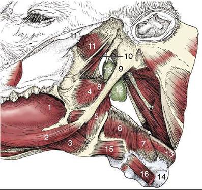
Figure 25-20 Connections of the pharynx and larynx with the base of the skull and the tongue. 1, Root of tongue; 2, styloglossus; 3, hyoglossus; 4, rostral pharyngeal constrictor; 5, middle pharyngeal constrictor; 6, 7, caudal pharyngeal constrictors (thyropharyngeus and cricopharyngeus); 8, stylopharyngeus caudalis; 9, stylohyoid; 10, tensor and levator veli palatini; 11, pterygoideus lateralis; 11', remnants of pterygoideus medialis; 12, medial retropharyngeal lymph node; 13, esophagus; 14, trachea; 15, thyrohyoideus; 16, sternothyroideus.