» The Pleura and the Lungs
The lungs are very unequal in that the right one is the larger by a ratio of 3:2. The asymmetry affects the disposition of the pleural sacs; the most obvious consequence is the deviation of both the cranial and the caudal mediastinum far to the left.
The cranial mediastinum actually attaches to the left wall of the thorax, while the caudal part meets the diaphragm in a sagittal plane that, when projected into the abdomen, bisects the reticulum, which exposes the two pleural sacs to almost equal chance of involvement when foreign bodies penetrate the thorax from that organ (p. 675). The apex of the right sac, which contains the tip of the cranial lobe of the lung, projects a few centimeters in front of the first rib, exposing it to risk of injury in penetrating wounds that are apparently confined to the base of the neck. The caudal reflection of the costal pleura onto the diaphragm is more important. It follows a cranially concave line that ascends steeply in its caudal part, tracing a course that passes through the 8th costochondral junction and the middle of the 11th rib before reaching the 12th rib just below the edge of the iliocostalis (see Fig. 27.2). Behind this line the diaphragm is directly attached to the thoracic wall, and the abdomen may be approached without risk of injury to the pleural sac. A space in front of this line, the costodiaphragmatic recess, is never fully exploited by the lung. Its extent may be considerably exaggerated after death, when the lung is collapsed.Apart from their asymmetry, the lungs of cattle are distinguished by their pronounced lobation and very evident lobulation.
The left lung possesses cranial and caudal lobes (see Fig. 27.2), and the former is divided into two parts: one extends forward toward the apex of the pleural sac, and the other descends ventrally over the pericardium. The notch between the two extends from the 3rd intercostal space to the 5th rib and defines the area in which the heart is in direct contact with the thoracic wall (Fig.
27.3). The basal border changes position with the phase of respiration; as a compromise between the inspiratory and expiratory positions, it may be described as following an almost straight line from the 6th costochondral joint to the upper part of the 11th rib. The thin marginal strip of lung does not provide useful clinical information, and the major area for percussion and auscultation is reduced to the surprisingly small triangle bounded by the triceps, the edge of the muscles of the back, and as the hypotenuse, the line joining the point of the elbow to the upper part of the 11th rib. A second (prescapular) area, extending a few centimeters in front of the ventral half of the cranial border of the scapula, is of minimal clinical significance.The right lung possesses four lobes—cranial, middle, caudal, and accessory (Fig. 27.4). The cranial lobe is independently ventilated by a bronchus detached from the trachea shortly before the bifurcation. The cardiac notch, smaller than that on the left side, is restricted to the lower parts of the 3rd and 4th spaces and is wholly under cover of the arm. The major area for clinical examination is a little larger on this side because it is free from the pressure exerted on the diaphragm by the rumen. Percussion toward the basal border is also more accurately performed because there is a sharp transition from the hollow lung sound to the duller note over the liver.
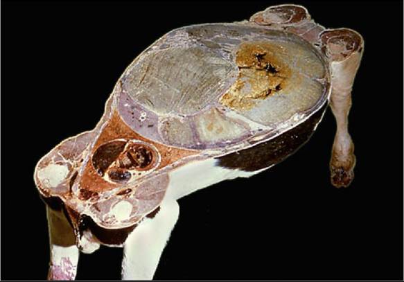
FIG. 27.1 Horizontal section at the level of the shoulder and stifle joints. Note the relative volumes of the thoracic and abdominal cavities.
Respiratory diseases cause significant economic losses to the cattle industry. Bronchial pneumonia occurs when infectious agents gain access to the lung through the airways. This occurs during physiologic stress induced by factors such as inclement weather and nutritional deficiencies, which lead to compromised immune defenses.
The common pathogens include bovine herpesvirus, Mycoplasma spp., and Mannheimia hemolytica. Thick connective tissue septa divide the lung substance and mark the surface where they impinge on the pulmonary pleura (see Fig. 4.27). These septa may help to localize infection, leading to a diseased part of the lung next to a normal part. This pattern is distinct from the diffuse pattern of the disease in the lungs of dogs.The capacity for respiratory exchange is limited by a relatively low total alveolar surface area and a lesser capillary density when compared with that of other species. A large part is required for basal needs, and little is held in reserve.
The lungs of the small ruminants are similar in gross form but show a lesser and usually patchy lobulation.
Although the circulation through the lungs is maintained by pulmonary and bronchial arteries, all the blood returns through a single set of veins. Two lymphatic plexuses drain the lungs. One lies directly below the pleura and drains this and the adjoining connective tissue. The other follows the peribronchial tracts and may be interrupted by the interposition of peribronchial nodes (though these are never conspicuous and cannot always be found). Both sets enter the tracheobronchial nodes placed about the origins of the principal bronchi.
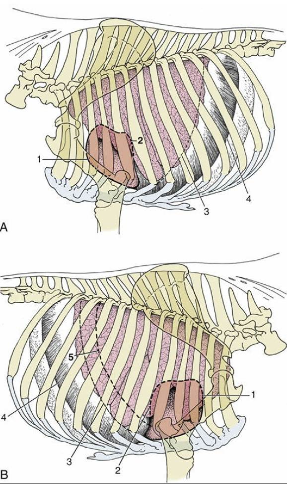
FIG. 27.2 (A) Left and (B) right projections of the bovine heart and lungs on the thoracic wall. The basal border of the lung and the line of pleural reflection are also shown. 1, Cranial extent of heart; 2, caudal extent of heart; 3, basal border of lung; 4, line of pleural reflection; 5, caudal border of lung percussion area, shown on right side.