The Proximal Tubule Has a High Capacity for H* Secretion and Bicarbonate Reabsorption
As described in Chapter 42, the proximal tubule normally reabsorbs the majority of the filtered HCO5-. The mechanism of bicarbonate reabsorption in the proximal tubule is illustrated in Figure 42-7.
In brief, apical membrane-bound carbonic anhydrase catalyzes the formation of H2O and CO2 from filtered HCO5- and secreted H*. The CO2 diffuses into the epithelial cell and combines with intracellular H2O under the influence of cytoplasmic carbonic anhydrase to form HCO3- and H+. HCO5 is transported across the basolateral plasma membrane by the Na*(HCO3-)5 co-transporter, driven by the electrical gradient for anions, and is reabsorbed into the blood. Concurrently, H* is transported into the lumen, primarily by the Na*∕H+ antiporter, but also by the H -ATPase pump, which may transport up to 35% of the total H' secreted by the proximal tubule. Thus, net HCO5 reabsorption and net H+ secretion are essentially equivalent terms in this system. The rate of bicarbonate reabsorption/acid secretion in the proximal tubule is increased by angiotensin II stimulation of the basolateral Na*(HCO5 )5 co-transporter, the Na+∕H+ exchanger, and the vacuolar H -ATPase.Although the proximal tubule has a large capacity for H* secretion (HCO5- reabsorption) and reabsorbs 80% to 90% of
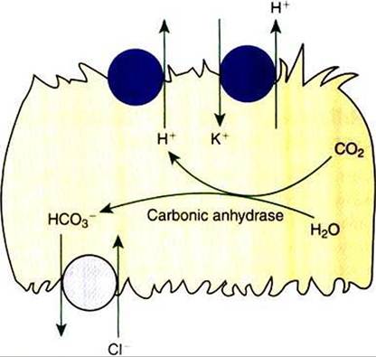
FIGURE 44-3 Schematic illustration of the mechanisms of H+ secretion and HCO3 reabsorption in the acid-secreting intercalated cells of the collecting duct.Two means of active transport of H’ across the apical plasma membrane are present: the electrogenic proton pump, H+-ATPaser and the electrically neutral H+,K+-ATPase pump.The intracellular formation of H* and HCO3 from CO2 and H2O is catalyzed by the enzyme cytoplasmic carbonic anhydrase.The basolateral plasma membrane contains a Cl /HCO3 exchanger that allows HCO3 reabsorption.
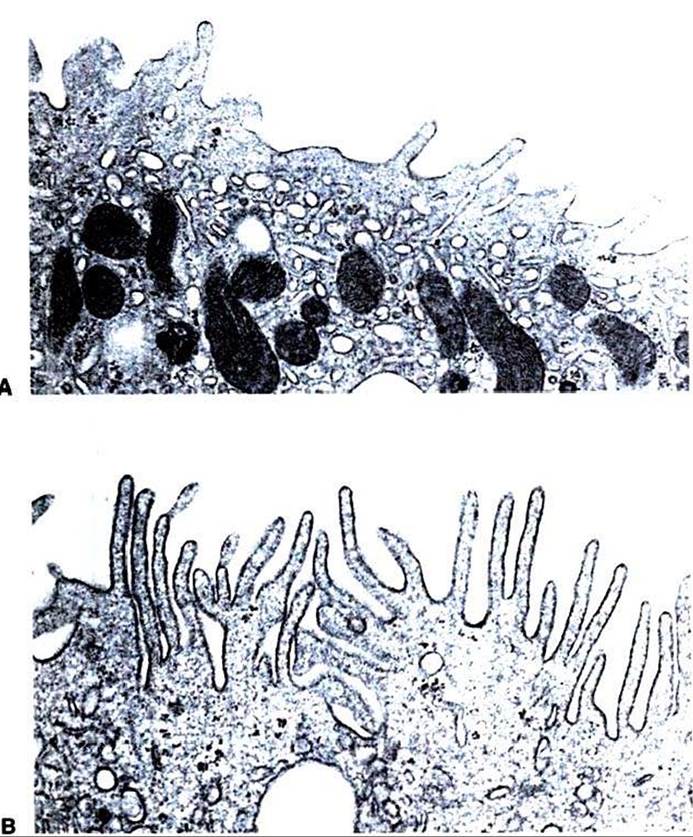
FIGURE 44-4 Transmission electron micrographs of an acid-secreting (type A) intercalated cell from rat cortical collecting duct.
A, In a control animal, the apical plasma membrane contains few small membranous projections, and the apical cytoplasm is filled with numerous membrane vesicles. B, In a rat with acute respiratory acidosis, the apical surface is covered with numerous long, membranous projections, and the number of apical cytoplasmic vesicular profiles is greatly reduced.This is the result of the insertion of membrane vesicles containing H* transporters into the apical plasma membrane in response to acidosis, thus enhancing the acid-secreting capacity of the cell. (Magnification ?11,300.)