The rather pendulous scrotum of the dog is globular and placed in a position intermediate between the perineum and the groin (Figure 15-17/11). It is most easily inspected from behind, and because it is sparsely
Guide to the Aging of Dog Fetuses
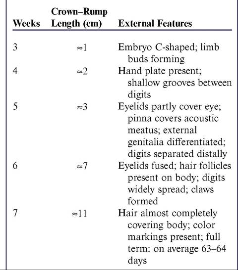
From Evans HE, Sack WO: Prenatal development of domestic and laboratory animals.
Growth curves, external features and selected references, Anat Histol Embryol 2:11-45, 1973.haired, its close molding on the testes is obvious. A deep groove defines the boundary between the internal compartments occupied by the generally asymmetrical testes. The thin scrotal skin and underlying fasciae do not impede palpation, which normally allows recognition of the body and tail of the epididymis, the deferent duct, and the spermatic cord, in addition to the testis itself. The scrotal skin of dogs is richly supplied with sweat glands. The scrotum of the cat is perineal, sessile, and commonly concealed by a dense covering of hair.
The testes are relatively small in both species. They are carried horizontally in dogs but with their caudal extremities tipped toward the anus in cats. Each testis is roughly oval in outline, laterally compressed, and related to the epididymis along its dorsal (in cats, craniodorsal) margin. The head and tail of the epididymis adhere to the testis, but the body is partly free, which creates a testicular bursa. The constituents of the compact spermatic cord disperse at the internal inguinal ring. Because of the very caudal position of the scrotum, the spermatic cord in the tom is unusually long. Perhaps it is because of this that the cremaster muscle of the cat is very weak. The striated cremaster muscle originates
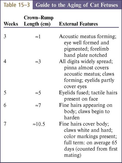
From Evans HE, Sack WO: Prenatal development of domestic and laboratory animals. Growth curves, external features and selected references. Anat Histol Embryol 2:11-45, 1973.
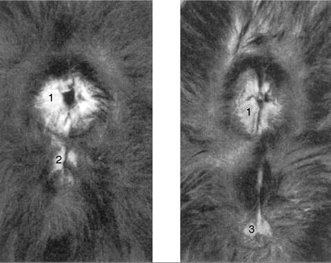
Figure 15-16 Perineum of 5-week-old littermate kittens. 1, Anus; 2, vulva; 3, prepuce.
from the iliac fascia on the ventral aspect of the psoas muscles just craniomedial to the caudal border of the internal oblique muscle, inserts on the internal spermatic fascia, and is innervated by the genitofemoral nerve.
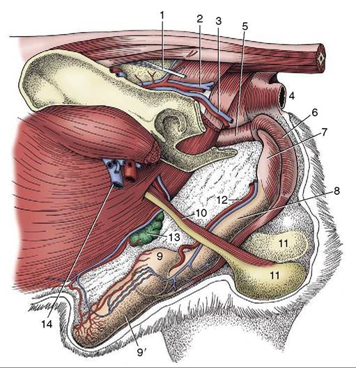
Figure 15-17 Deep dissection of the external reproductive organs of the dog. 1, Sacrotuberous ligament; 2, caudal gluteal vessels; 3, internal pudendal vessels; 4, anus; 5, pelvic urethra; 6, bulb of penis enclosed by bulbospongiosus; 7, ischiocavernosus over left crus; 8, body of penis; 9, 9', bulbus and pars longa glandis; 10, spermatic cord; 11, testes in scrotum; 12, dorsal artery and vein of the penis; 13, superficial inguinal lymph nodes and caudal superficial epigastric vessels; 14, femoral vessels.