THE RECTUM AND ANUS
Although the origin of the rectum is arbitrarily defined, its most caudal part is distinguished from the colon by a wider caliber and more muscular wall. The interior, marked by impermanent transverse folds, is generally distended with feces (Figure 29-6).
The colic mesentery continues as the mesorectum, which abruptly shortens to a mere 3 cm, before gradu- The bladder is intraabdominal in the young calf. In the adult it is confined to the pelvic cavity when empty but extends forward over the abdominal floor when distended. The neck within the pelvis is without a peritoneal covering and is attached to the pelvic floor by fat and loose connective tissue (see Figures 29-7 and 29-8). Urine escaping from a ruptured bladder, which is a relatively common mishap, especially in steers, may infiltrate this tissue or enter the peritoneal cavity according to the site of the tear. There are the usual lateral and median ligaments.
The relations of the bladder naturally vary. In the cow, it is always in contact with the cranial part of the vagina and the cervix and often with the body and horns of the uterus. Within the abdomen it makes contact with the dorsocaudal blind sac of the rumen and with the intestines (Figure 29-11).
The urethra is much narrower than that of the mare and runs below the vagina, to which it becomes increasingly attached as it proceeds caudally. It opens into the vestibule through a median slit that is shared with the suburethral diverticulum (Figure 29-11/73), a blind pouch extending cranially that is large enough to admit the end joint of a finger. The pouch can be a nuisance when catheterization is attempted. The urethralis muscle only covers the caudal part of the urethra, which more
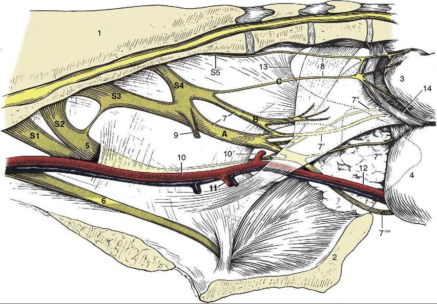
Figure 29-5 Nerves and vessels on the medial surface of the bovine pelvic wall. Local anesthesia of the pudendal nerve can be obtained by injections at A and B; anesthesia of the caudal rectal nerves is possible by an injection at C. 1, Sacrum; 2, pelvic symphysis; 3, rectum (reflected); 4, vagina (reflected); 5, sciatic n.; 6, obturator n.; 7, pudendal n.; 7, distal cutaneous branch of pudendal n.; 7", proximal cutaneous branch of pudendal n.; 7"', deep perineal n.; 7'"', continuation of pudendal n. to clitoris; 8, caudal rectal nn.; 9, pelvic n.; 10, internal iliac a.; 10', caudal gluteal a.; 11, vaginal a.; 12, internal pudendal a.; 13, caudal border of sacrosciatic ligament; 14, retractor clitoridis.
cranially is anchored to the floor by a short but strong ligament. The cranial fascicules of the urethralis muscle insert on a dorsal raphe that completes the encirclement of the urethra; the more caudal ones form a U that attaches to each side of the vagina and vestibule, enclosing both the diverticulum and the urethra.
The blood supply to these organs comes from the umbilical and vaginal arteries.