THE REPRODUCTIVE TRACT DURING PREGNANCY
The ovaries continue to show cyclical activity during the first months of pregnancy. Although the first corpus luteum does not persist beyond the usual term, it is replaced by a succession of other corpora over the next 5 months; some are formed after rupture of follicles, others apparently by direct luteinization.
The accessory corpora lutea survive longer than the original one and are a rich source of progesterone. The growth, ripening, and luteinization of the new follicles are controlled by gonadotrophic hormones derived from the endometrial cups that are so distinctive a feature of the species. After 5 months the accessory corpora lutea also regress and pregnancy is then maintained by progesterone of placental origin. The enormous enlargement of the fetal gonads, peculiar to the horse among domestic species, reaches a peak between 6 and 8 months. Despite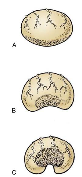
Figure 22-15 The postnatal development of the ovary. The more rapid growth at the poles confines the germinal epithelium (stippled) to a small central area. A, At birth; the germinal epithelium is widespread over the surface. B, At 6 months of age. C, Adult; the germinal epithelium surrounds an indentation known as the ovulation fossa.
assertions that fetal hypophysial luteinizing hormone (LH) is responsible for the enlargement, unpublished information indicates that the enlargement continues in the decapitated fetus; this points to the endometrial gonadotrophins as a contributing if not sole source (see Figure 22-18, B and see p. 576). The temporary enlargement of the fetal testes influences the timing and the success of their descent, which is normally completed about full term.
The proliferative changes of the endometrium that occur with each cycle continue and intensify if pregnancy has occurred.
The early diagnosis of pregnancy and, because of the prevalence of early embryonic death, the confirmation of its continuation through the critical early stages have particular importance in equine practice. An additional significance is provided by the desirability of recognizing twin pregnancies at an early stage. Twin pregnancies are rarely completed successfully, and the clinician and client may choose to destruct one of the twins by manual crushing to lessen the risk of losing a breeding season. The crushing has to be carried out before implantation. Although various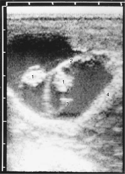
Figure 22-16 Ultrasonographic view of 31-day equine twin embryos. The scale is in centimeters. 1, Twin embryos; 2, junction of the two conceptuses; 3, developing allantoic membrane; 4, uterine wall.
laboratory methods of pregnancy diagnosis exist, the principal reliance remains on careful internal examination per rectum, supplemented by ultrasonography (Figure 22-16). The experienced clinician may recognize a loss of uterine tone at the location of a conceptus, compared with the tone of neighboring parts, as early as the 20th day—possibly even a day or two before this. The location of the conceptus at this time is within the part of a uterine horn adjacent to the junction with the body of the organ; at this stage the conceptus has a diameter of approximately 30 to 40 mm, and a slight bulge of the ventral aspect of the gravid horn should be detectable. Ultrasonic examination may bring forward the time of recognition of the presence of a conceptus to as early as the 11th or 12th day, occasionally even the 9th day. Because the conceptus has a diameter of only a few millimeters at this stage, it is clear that very systematic examination is required to detect, or confidently exclude, its presence. The identification of the body of the embryo becomes possible a week or so later (about day 19), and this removes any lingering suspicion that a cavity identified at an earlier examination might be attributable to an endometrial cyst.
The differentiation of pregnancy from pathology will probably receive additional confirmation from a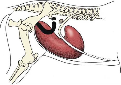
Figure 22-17 Changes in the topography of the uterus and ovary between the beginning (black) and the end (red) of pregnancy.
shift in location of the conceptus, which is still mobile, unlike a lesion.
The early conceptus enjoys considerable mobility before adopting a fixed location within the uterus. There is evidence to suggest that, though most equine conceptuses are located within the body of the uterus about the 10th day, they will have settled within a horn a week or so later.
Ultrasonography may be employed at a somewhat later stage of pregnancy to determine the sex of the fetus, which is revealed by the location of the genital tubercle; it is found close to the umbilical cord in the male, nearer the tail in the female. Such examinations are performed after 55 days.
The whole gravid horn (which is more commonly the right one) then gradually enlarges, followed by the body and, although to a lesser degree, the nonpregnant horn. As the uterus enlarges it sinks into the abdomen, dragging the body and the cervix out of the pelvis (Figure 22-17). The broad ligaments exert constraint on the mesometrial margins, and the horns therefore enlarge asymmetrically and become more flexed on themselves; the ovaries are drawn ventrocranially. The uterine arteries, which are pulled in the same direction, develop a characteristic vibration (fremitus or thrill) in the pregnant mare. This feature may be appreciated on rectal examination, and its diagnostic value is greatest at that stage of pregnancy (between the 3rd and 5th month) when the uterus has sunk out of reach. The position of the foal adapts to the form of the uterus; by midpregnancy it has come to lie with its back against the greater curvature of the horn (and thus ventrally) and with its head generally (99% of the time) raised toward the cervix.
In the circumstances that most favor easy parturition, the bulky body of the foal is preceded into the cervix by the extended forelimbs, on which rest the relatively small head and slender neck. The foal is delivered with its back uppermost. Because of the general enlargement and considerable size of the body of the uterus, it is possible for the occasional fetus to lie transversely, extending from one horn into the other; clearly this bodes ill for parturition. Enlargement of the uterus displaces the other abdominal contents forward and upward; in later pregnancy the uterus dominates the entire abdominal topography, extending forward on the abdominal floor and under the rib cage; however, it generally remains to the left of the cecum.A prominent feature of the uterus in early months of pregnancy is the presence of a ring or horseshoe formation of scablike structures, disfiguring the endometrium of the caudal part of the horn, the location where the young conceptus comes to rest. These so- called endometrial cups (Figure 22-18, B) are unique to Equidae and are the source of both equine chorionic gonadotropin (formerly known as pregnant mare’s serum gonadotropin [PMSG]), the hormone responsible for the unusual activity of the ovary of the pregnant mare (p. 574) and the even more remarkable, though
temporary, enlargement of the gonads of equine fetuses of both sexes. The cups have their origin in cells that invade the endometrium from a limited region of the chorion: the (allanto-) chorionic girdle that marks the boundary between the allantochorionic and omphalo- chorionic (yolk sac) portions of the embryonic vesicle and provides the area of initial adhesion of the conceptus to the uterus (Figure 22-18, A). The migration of chorionic cells begins about the 35th day, and the cups soon become visible as low endometrial elevations. They continue to grow, forming irregular centrally depressed saliences that reach their zenith about the 60th day, only to enter a process of degeneration and necrosis shortly thereafter.
The process culminates in their separation and sloughing from the endometrium, which are events largely concluded by the 120th day (or thereabout), although a few may persist much longer. The fetal (chorionic) cells penetrate some way into the endometrial stroma, and although they provide the essential endocrine components of the cups, they become admixed with connective tissue cells, blood vessels, and glandular debris and secretion contributed by the endometrium. Some detached cups come to lie between the endometrium and chorion; other detachments of this material push into the allantoic cavity, enclosed within pedunculated sacs of allantochorion, and these protrusions may be the origin of some of the hippomanes mentioned shortly.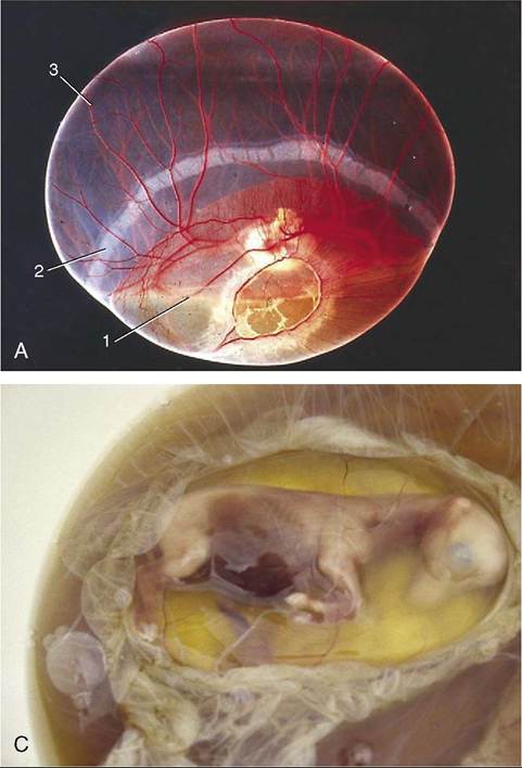
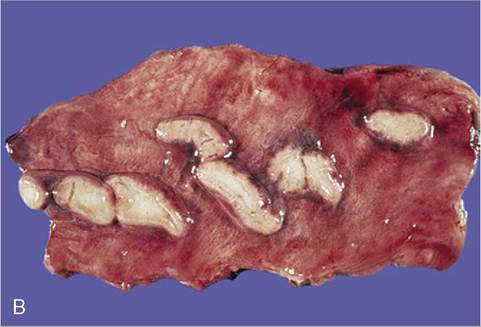
Figure 22-18 A, Young conceptus (horse). 1, Yolk sac; 2, chorionic girdle; 3, allantochorion. B, Endometrial cups (mare) during early pregnancy. C, The placenta of the horse fetus is not very complex. The villi do not penetrate deep into the endometrium.
The cervix of the pregnant mare is firm and closed by a plug of mucus (see Figure 22-12). The pale vaginal wall is also coated with mucus that becomes stickier and more inspissated as pregnancy progresses. The connective tissues of the cervix, vagina, and vulva and the sacrotuberal ligaments soften shortly before birth, which is generally speedily executed, facilitated by the generous dimensions of the pelvic cavity. It is necessary that it should be so, as rupture of the membranes with loss of fetal fluids allows separation of the loose attachment between the chorion and the endometrium, jeopardizing fetal respiration.
Puerperal changes follow the same pattern as in other species but run a rapid course. Involution of the uterus is completed sooner than in the cow, and because there is no endometrial damage to repair, mares covered at the “foal heat”—about the 8th to 10th day after giving birth—often conceive.