THE ROOF OF THE ABDOMEN
The bodies of the lumbar vertebrae, the sublumbar muscles, and the diaphragm furnish the roof of the abdomen. The aorta and caudal vena cava lie within the cleft between the two psoas minor muscles: the artery is to the left, and the vein is to the right (Figure 21-22/6,7).
The branches of the aorta and the tributaries of the vein are, in principle, the same as in other species.The autonomic nerves and ganglia show some specifically equine features, although these are matters of detail rather than of substance. The general pattern is shown in Figure 21-24. The fused celiac and cranial mesenteric ganglia lie ventral to the aorta, to each side of the celiac and cranial mesenteric arteries. The right and left ganglia are joined by bridges cranial and caudal to the latter artery. They are sizable structures, 5 cm or so long, and are generally unequal: the left complex is larger and more regular than the right one (Figure 2124/75). Each is joined by a major splanchnic nerve and, in varying fashion, by parasympathetic fibers from the dorsal vagal trunk. The nerves that leave the ganglia follow the arteries, branching where these branch and forming a dense plexus in which the sympathetic and parasympathetic contributions mingle. The whole plexi- form arrangement that radiates from the major ganglia is known as the celiacomesenteric (solar) plexus. Additional small renal ganglia occur on the nerves about the renal arteries.
The celiacomesenteric complex is joined to the caudal mesenteric plexus by a plexus on the aorta and an additional trunk that runs at a more ventral level within the colic mesentery.
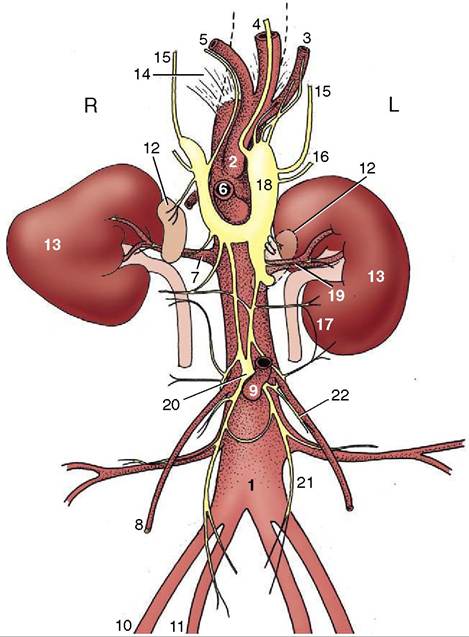
Figure 21-24 Schema of the abdominal autonomic nerves and branches of the abdominal aorta, ventral view. 1, Aorta; 2, celiac a.; 3, splenic a.; 4, left gastric a.; 5, hepatic a.; 6, cranial mesenteric a.; 7, renal a.; 8, testicular (ovarian) a.; 9, caudal mesenteric a.; 10, external iliac a.; 11, internal iliac a.; 12, adrenal glands; 13, kidneys; 14, crus of diaphragm; 15, major splanchnic nn.; 16, minor splanchnic nn.; 17, lumbar splanchnic nn.; 18, combined celiac and cranial mesenteric ganglia; 19, renal plexus; 20, caudal mesenteric ganglion; 21, hypogastric n.; 22, testicular (ovarian) plexus.
The caudal mesenteric ganglion lies cranial to the origin of the like-named artery (Figure 21-24/9,20). It gives rise to nerve plexuses that follow this vessel and the gonadal vessels to the small colon and reproductive organs, respectively, and to the hypogastric nerves (Figure 21-24/27) that pursue a retroperitoneal course on the roof of the pelvis. Lumbar splanchnic nerves join the major ganglia and the aortic plexus in an erratic fashion.
The usual direct detachment of preganglionic fibers from the splanchnic nerves to the medullary parts of the adrenal glands exists.